
Scientific research on spruce resin at a glance
- The strongest laboratory evidence concerns antimicrobial activity and biofilms. Materials derived from Norway spruce resin (Picea abies) have inhibited various microorganisms in several experimental systems, although the result depends strongly on concentration, contact time and formulation.
- The research extends well beyond bacteria. Studies have examined dermatophytes and other fungi, keratinocytes, fibroblasts, endothelial cells, as well as individual resin acids and other compounds present in resin materials.
- Human clinical data also exist. Resin preparations have been studied in chronic and surgical wounds, although clinical studies are far fewer than laboratory studies and have important methodological limitations.
- Not all studies investigated the same material. Natural spruce resin, aged resin, different resin preparations and isolated compounds are not interchangeable. For each finding, we therefore make clear what was actually tested.
- The overall research picture is therefore multi-layered. Current findings point to several possible biological pathways – from direct effects on microorganisms to processes in tissue cells – but they cannot fairly be reduced to a single “active ingredient” or a single mechanism.
Table of contents
What did researchers actually study?
When interpreting research on spruce resin, one of the most important questions is often very simple: what was actually tested? Resin is a naturally heterogeneous mixture, and its composition changes with the type of resin secretion, the age of the material and the way the sample is prepared. A finding from one resin fraction or one isolated compound is therefore not automatically a finding about “spruce resin” as a whole.
| What was studied | What such a study can tell us | Important limitation |
|---|---|---|
| Natural Norway spruce resin (Picea abies) | Provides the most direct evidence about the chemistry and biological properties of spruce resin as a natural material. | Natural resin itself is not uniform: fresh exudate, aged resin and resin associated with different types of tree injury may differ chemically. |
| Aged resin and emulsions or extracts prepared from it | These systems allow researchers to study less volatile constituents, antimicrobial activity, biofilms and other properties of aged resin material. | The preparation of the sample and the availability of individual compounds within the formulation can strongly influence the result. |
| Individual resin acids and other isolated compounds | They help identify which molecules may contribute to a particular biological effect and which cellular pathways may be involved. | An effect of an isolated compound does not mean that this compound alone explains the activity of complex natural resin. |
| Resins and resin compounds from other conifers | They can add to our understanding of shared terpenoid and resin-acid mechanisms in related natural materials. | Such findings cannot be directly attributed to Picea abies resin. |
| Resin preparations used in clinical studies | They show what happened in actual patients under a defined study protocol, for example when wound healing was monitored. | The result applies to the preparation and protocol that were studied and cannot automatically be transferred to every product containing spruce resin. |
For this reason, in the sections below we distinguish between the test material, the type of study and the outcome that was actually measured. This makes it possible to judge more accurately what is well established and where conclusions remain indirect.
Antimicrobial activity of spruce resin
Antimicrobial activity is currently one of the best-studied biological properties of resin materials. Different research groups have shown that Norway spruce (Picea abies) resin can inhibit bacteria and, in some experimental systems, yeasts as well. Importantly, the outcome depends strongly on the concentration of resin material, contact time, solubility and formulation.
Early direct evidence from Norway spruce resin
In 2007, Rautio and colleagues studied natural Picea abies resin and a traditional resin salve. In laboratory tests, the resin inhibited the growth of all tested Gram-positive bacteria, including Staphylococcus aureus, MRSA, Staphylococcus epidermidis, Enterococcus faecalis, VRE and streptococci.
The effect was considerably weaker against most of the Gram-negative bacteria tested in this system. When the bacteria were transferred to resin-free growth media after exposure, they were able to grow again. The effect was therefore described primarily as bacteriostatic: the resin inhibited bacterial growth but did not necessarily kill the cells under the conditions used.
A different test revealed a broader antimicrobial spectrum
In 2011, Sipponen and Laitinen tested purified resin material obtained from Picea abies trunks using the European Pharmacopoeia challenge test. At a concentration of 10% in an inert salve base, the numbers of S. aureus and MRSA were significantly reduced within 24 hours. Over a longer exposure period, reductions were also observed for Escherichia coli, Pseudomonas aeruginosa, Bacillus subtilis and the yeast Candida albicans.
This is important because it shows that the earlier weak result against Gram-negative bacteria did not necessarily mean that these organisms were inherently resistant to resin material. Resin compounds are poorly soluble in water, and agar diffusion tests can therefore substantially underestimate their activity. With greater concentration and direct contact, the observed spectrum can be considerably broader.
Aged spruce resin and water-accessible resin acids
Haapakorva and colleagues later investigated emulsions prepared from aged Picea abies resin material. In this system they also found pronounced antimicrobial activity against Gram-positive and Gram-negative bacteria, MRSA and yeasts.
The chemistry of the water-accessible fraction was particularly interesting. Oxidised resin acids were present, whereas the major non-oxidised resin acids were not detected in the aqueous phase. This supported the view that oxidised derivatives may play an important role in this type of aged, water-accessible system.
An important addition: non-oxidised abietic acid is active as well
More recent research has shown that the picture cannot be limited to oxidised resin acids alone. In 2020, Ito and colleagues demonstrated that isolated abietic acid inhibited the growth of Streptococcus mutans and the formation of its biofilm.
In 2026, Park and Lee tested isolated abietic acid against S. aureus, P. aeruginosa, E. coli and C. albicans. Under laboratory conditions, it inhibited the growth of all four microorganisms. These two studies used abietic acid derived from pine resin material and are therefore not direct studies of spruce resin. They do, however, demonstrate that one of the resin acids found in conifer resins is biologically active in its own right.
Overall picture: the direct antimicrobial activity of resin materials cannot reasonably be attributed to a single compound. Current evidence better supports a broader model in which parent resin acids and their oxidised derivatives may both contribute, while the observed effect depends on chemical profile, concentration, solubility, formulation and the microorganism involved.
Biofilms: preventing formation is not the same as acting on an established biofilm
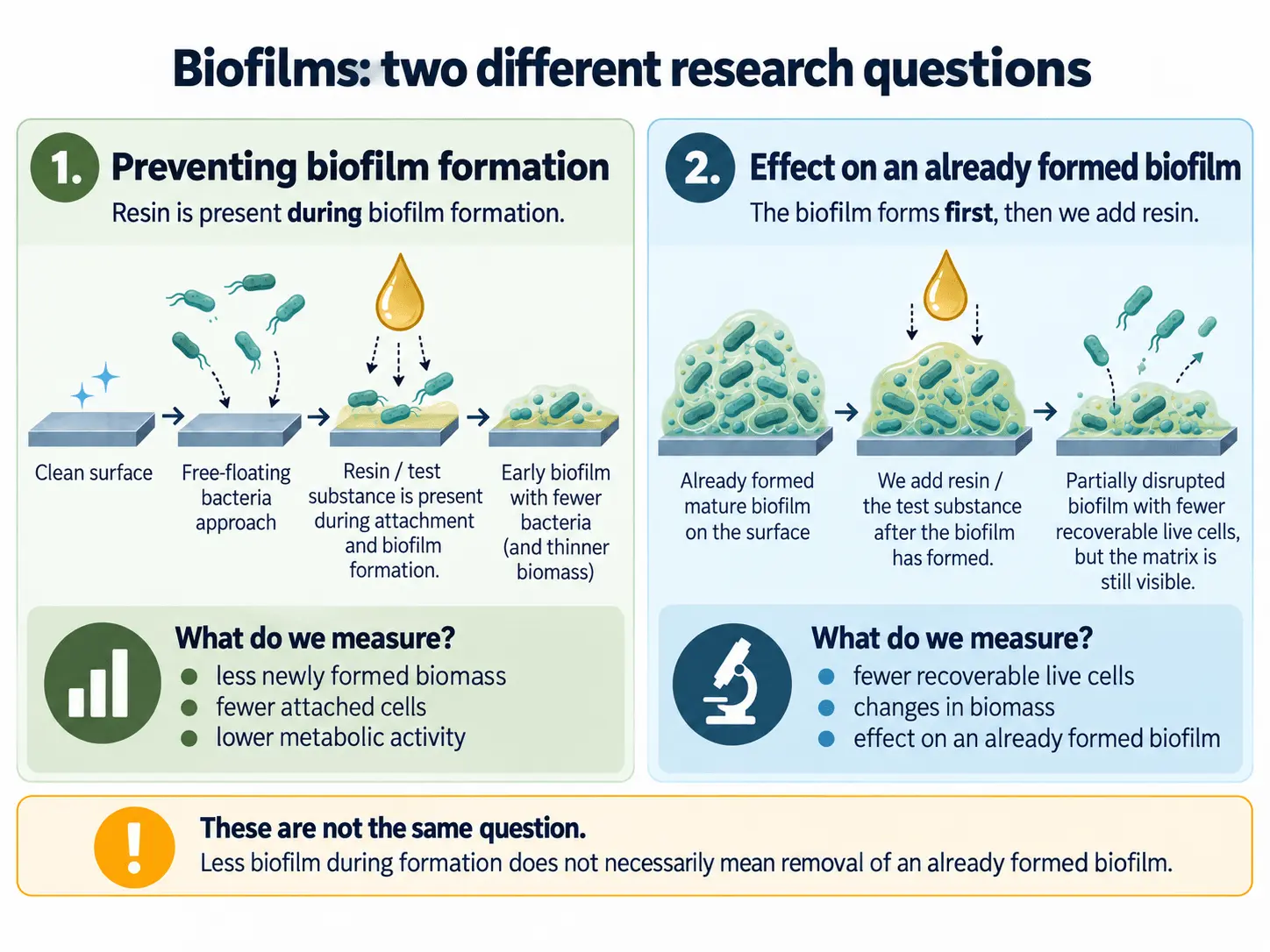
A biofilm is not simply a collection of freely floating microorganisms. Bacteria or fungi attach to a surface, form an organised community and surround themselves with a matrix that alters their metabolism and the accessibility of antimicrobial substances. Research must therefore distinguish between inhibiting biofilm formation and acting on a biofilm that was already established before exposure.
Aged spruce resin also affected an already established staphylococcal biofilm
In 2017, Haapakorva and colleagues studied an emulsion prepared from aged Norway spruce (Picea abies) resin material. They used MRSA Staphylococcus aureus and Staphylococcus epidermidis on medical silicone discs in a laboratory model designed to mimic some features of the chronic-wound environment.
In one experiment, the resin emulsion was present while the biofilm was forming. In another, the bacteria were first allowed to develop a biofilm for 24 hours before the test preparation was added. In both settings, the spruce resin emulsion significantly reduced the number of recoverable staphylococcal cells.
This is a more specific finding than merely observing reduced biofilm biomass during formation. However, it should not be described as complete “biofilm removal”: the researchers measured viable or recoverable bacterial cells after mechanical removal from the disc, not complete disappearance of the biofilm matrix or sterilisation of the surface.
Abietic acid also showed antibiofilm activity on its own
In 2020, Ito and colleagues showed that isolated abietic acid reduced Streptococcus mutans biofilm formation, lowered biofilm metabolic activity and reduced recoverable cells after short-term exposure of an already established biofilm.
In 2026, Park and Lee extended the findings to several other microorganisms. Isolated abietic acid reduced biofilm biomass in a concentration-dependent manner for S. aureus, Pseudomonas aeruginosa, Escherichia coli and Candida albicans. In this study, however, abietic acid was present during biofilm formation, so the result does not demonstrate removal of mature biofilms of these microorganisms.
What can we conclude from these studies?
Taken together, the findings indicate that resin materials and individual resin acids can affect several different biofilm-related processes. Of particular importance is the direct laboratory evidence that aged spruce resin material can reduce surviving bacterial cells in an already established staphylococcal biofilm.
It has not yet been demonstrated, however, that biofilm effects are the main reason for the clinical effects of resin preparations in wounds, nor can the same activity be attributed to a specific Smrekovit product without direct testing.
Fungi and dermatophytes: the effect depends on the species and test system
It is not useful to speak of a single universal “antifungal effect”. Dermatophytes, Candida yeasts and other opportunistic fungi differ substantially in their biology, so a result against one group cannot automatically be transferred to another.
Norway spruce resin strongly inhibited dermatophytes
In 2011, Rautio and colleagues studied purified Norway spruce (Picea abies) resin against dermatophytes, fungi adapted to growth on keratinised tissues. Species from the genera Trichophyton, Microsporum and Epidermophyton were tested.
Zones of inhibition were observed for all dermatophytes tested. Among Trichophyton species, the effect was concentration-dependent: weaker at lower resin concentrations and more pronounced at higher ones.
Microscopy revealed direct damage to fungal hyphae
In Trichophyton mentagrophytes, electron microscopy showed deformed, twisted and irregular hyphae together with damage to the fungal cell envelope. Dead or disrupted fungal structures were present within the inhibition zone, while normal hyphae were found outside it.
This provides relatively direct morphological evidence that the resin material did more than simply slow fungal growth. It does not, however, identify which individual compound in the complex resin was responsible or define a single molecular target.
With Candida, the test method substantially changed the result
In the same study, activity against several Candida species was very weak or absent in agar diffusion tests. Later studies produced a very different picture when the resin material was placed in more direct contact with the microorganism.
Sipponen and Laitinen showed a concentration- and time-dependent reduction of recoverable Candida albicans cells in a contact challenge test. Haapakorva and colleagues also observed pronounced activity against C. albicans and C. tropicalis using an aqueous emulsion prepared from aged spruce resin material.
The difference between these results is very likely related to a key property of resin acids: their poor water solubility. In an agar diffusion assay, an active compound must first diffuse through an aqueous environment, which can substantially underestimate the activity of lipophilic resin material.
Abietic acid itself inhibits Candida albicans
In 2026, Park and Lee tested isolated abietic acid derived from pine resin material against C. albicans. In a liquid laboratory system, the minimum inhibitory concentration was 62 µg/ml and the minimum fungicidal concentration was 500 µg/ml.
This means that a substantially higher concentration was required to prevent recoverable fungal growth completely than to inhibit visible growth. Importantly, this was not a study of spruce resin, but of an isolated compound.
The antifungal spectrum is not universal
The evidence does not support the claim that resin materials act against all fungi. In key experiments, opportunistic fungi including Fusarium solani, Chrysosporium keratinophilum and Aspergillus brasiliensis were poorly susceptible or not susceptible to the resin systems used.
The most defensible current conclusion is therefore quite specific: the strongest direct antifungal signal for Norway spruce resin is against dermatophytes, while activity against Candida depends strongly on formulation, solubility, concentration and contact time.
Keratinocytes and re-epithelialisation: what did studies on skin cells show?
In addition to microorganisms, researchers have also examined the direct effects of resin materials on skin cells. For spruce resin, the 2020 study by Goels and colleagues is particularly important because it linked the chemical composition of Picea abies material with a laboratory keratinocyte assay within the same study.
What did the researchers actually measure?
They used HaCaT cells, a widely used human keratinocyte cell line. A standardised cell-free gap was created in a uniform cell layer, and the researchers measured how much of this area became covered by cells again over 24 hours.
This gap-closure assay is a laboratory model of a process associated with re-epithelialisation. However, it does not distinguish precisely how much closure results from cell migration and how much from proliferation. The result is therefore not equivalent to direct evidence of faster wound healing in humans.
Non-volatile fractions of the spruce material showed the main activity
The researchers separated the spruce balm into several chemical fractions and tested them individually. Two fractions significantly increased gap closure: the acidic E1 fraction by approximately 47% compared with the vehicle control, and the apolar E3 fraction by approximately 36%.
A negative result is also informative: the essential oil obtained from the same spruce material did not increase gap closure in this assay. This does not mean that volatile compounds have no other biological effects, but it shows that they were not the main drivers of the response in this particular keratinocyte model.
Which individual compounds showed an effect?
Positive signals were observed for dehydroabietic acid (DHAA), pimaric and isopimaric acids, several hydroxylated DHAA derivatives and the lignan pinoresinol. Some combinations of isolated compounds produced a stronger response than the individual compounds alone.
This is important because it argues against a simple explanation in which one single “active ingredient” accounts for the biological properties of a complex resin. Activity was distributed among several chemically different non-volatile constituents.
The whole spruce balm did not produce a statistically significant result in this assay
An important detail can easily be overlooked. At the tested concentration, the whole purified spruce balm showed an approximately 14% trend towards greater gap closure, but this result was not statistically significant. Stronger and statistically significant signals appeared only after individual fractions and compounds were tested.
This illustrates why it is important to distinguish between whole resin material, a particular fraction and an isolated compound. A result observed at one level cannot automatically be attributed to another.
Resin does not always “stimulate” keratinocytes in the same direction
Other experimental models further show that the keratinocyte response should not simply be described as stimulation. In a psoriasis-like mouse model, processed resin material reduced markers of pathological epidermal hyperproliferation, whereas the Goels model of an interrupted cell layer showed increased gap closure.
These findings are not necessarily contradictory. Re-covering a defect is desirable in an injured surface, whereas reducing excessive proliferation can be desirable in pathologically hyperproliferative skin. It is therefore more accurate at present to speak of a context-dependent influence of resin compounds on the epidermal response rather than a general “stimulation of keratinocytes”.
Fibroblasts and extracellular matrix: what happens in an inflammatory environment?
Fibroblasts have a different role from keratinocytes in repair of damaged tissue. They participate in migration into the injured area, formation and organisation of the extracellular matrix and remodelling of newly formed tissue. Importantly, the goal is not simply “more fibroblast activity”: an excessive or prolonged response may also contribute to excessive scarring.
Dehydroabietic acid was tested on human dermal fibroblasts
In 2014, Wang and colleagues used human adult dermal fibroblasts and added TNF-α to create a simplified laboratory model of an inflammatory environment in which normal reparative cell functions are impaired.
They tested isolated dehydroabietic acid (DHAA), one of the diterpene resin acids that has also been directly identified in Norway spruce (Picea abies) material.
DHAA mitigated several adverse effects of TNF-α
Added TNF-α reduced fibroblast proliferation, increased apoptotic responses and impaired several processes associated with reparative cell function. DHAA partially shifted several of these changes back towards control values.
Among other effects, DHAA mitigated TNF-α-induced suppression of proliferation, reduced apoptotic signalling and improved the migratory response of fibroblasts under the appropriate experimental conditions.
An important detail is that the study did not show that DHAA lowers TNF-α concentrations. TNF-α was added externally to the fibroblasts. The study showed that DHAA altered the way fibroblasts responded to an already present inflammatory signal.
Effects were also observed in TGF-β1/Smad signalling and matrix-related responses
TNF-α disrupted TGF-β1/Smad3 signalling in this model, a pathway involved in fibroblast reparative processes. DHAA partially mitigated this inhibitory effect and preserved more of the TGF-β1 response.
Under the appropriate conditions, expression of α-SMA and fibronectin was also increased or restored. The former is associated with the contractile myofibroblast phenotype, whereas the latter is an actual component of the extracellular matrix.
This does not mean that DHAA made the entire formation of new matrix “better”. The study did not directly measure collagen organisation, mechanical quality of newly formed tissue, long-term remodelling or the balance between repair and fibrosis.
Complex resin materials may influence fibroblasts in other ways as well
In 2007, Seo and Suk studied several traditional Resina Pini preparations on human gingival fibroblasts. Some preparations reduced prostaglandin E2 (PGE2) production, with substantial differences depending on processing and the fraction used.
This was not a study of spruce resin and it did not measure migration, TGF-β/Smad signalling or extracellular matrix remodelling. It does, however, demonstrate an important point: the fibroblast response can depend strongly on the composition and preparation of the resin material.
What does this mean for our understanding of spruce resin?
DHAA is one of the more interesting molecular bridges between the chemistry of spruce resin and cellular processes: it has been confirmed as a constituent of spruce balm and, in separate laboratory models, has shown effects on both keratinocytes and dermal fibroblasts.
This still does not mean that whole natural resin or a specific Smrekovit product affects fibroblasts in the same way as isolated DHAA. Such a conclusion would require direct testing of a chemically characterised sample of the actual resin on human dermal fibroblasts.
The historical context of resin use in wounds is presented separately on our page about traditional use of spruce resin; laboratory cell studies help us identify which biological pathways may be worth investigating further.
Angiogenesis: can resin acids influence the formation of new blood vessels?
During wound healing, newly forming tissue requires oxygen and nutrients, making angiogenesis – the formation of new blood vessels from existing vasculature – an important part of the proliferative phase. Among resin compounds, abietic acid has been studied directly in this context. It is one of the diterpene resin acids also found in Norway spruce resin.
Abietic acid affected human endothelial cells
In 2017, Park and colleagues used human umbilical vein endothelial cells (HUVECs) to study isolated abietic acid and pine resin material. They measured cell migration, organisation into tube-like structures and closure of an experimentally created cell gap.
At 0.8 µM abietic acid, formation of tube-like structures increased by approximately 24% compared with the control, while HUVEC migration increased by approximately 25%. These effects were observed at low concentrations that did not significantly reduce cell viability.
Tube-like structures are not yet functional new blood vessels
Such laboratory assays are useful for assessing angiogenic potential, but they have an important limitation. Endothelial cells organising into network-like or tube-like structures on a matrix does not mean that functional, perfused blood vessels have formed in living tissue.
HUVECs are also not adult human dermal microvascular endothelial cells. The study therefore demonstrates an interesting endothelial signal rather than direct evidence of new vessel formation in a human wound.
ERK and p38 were activated, but their causal role was not confirmed
Exposure to abietic acid increased phosphorylation of the ERK and p38 signalling pathways. However, inhibitors of these pathways did not clearly abolish the increased tube formation or migration.
It would therefore be incorrect to state that abietic acid causes angiogenesis through ERK or p38. The study shows that these signals were activated alongside the observed effects, not that they were proven causal mediators.
Wounds closed faster in a mouse model
The same research group also tested abietic acid in a mouse skin-wound model. The abietic-acid group showed faster reduction in wound area than the control group at several time points.
Importantly, the study did not directly measure new-vessel density, endothelial markers or functional perfusion in the wound. Faster wound closure therefore cannot be taken as proof that angiogenesis caused the effect.
The evidence base remains much narrower than for antimicrobial activity
The Park study is interesting, but the current research collection does not contain a new independent study directly replicating the angiogenic effect of abietic acid. In addition, the abietic acid used was derived from pine resin material rather than spruce resin.
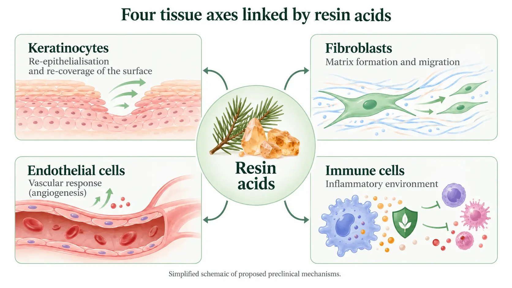
The most accurate current conclusion is therefore that abietic acid affects several endothelial processes associated with angiogenesis in one preclinical research system. This supports the possibility that non-volatile resin acids may also influence the vascular component of tissue repair, but this has not been demonstrated directly for whole spruce resin – and even less so for a specific Smrekovit product.
Inflammatory response and cytokines: resin acids do not always act in the same direction
Inflammation is a normal part of the response to tissue injury. During the early phase, cytokines such as TNF-α, IL-1 and IL-6 are important for activating immune cells and initiating tissue repair. Problems arise mainly when the response is excessive, prolonged or poorly timed.
Research on spruce resin and resin acids therefore cannot be reduced to asking whether a substance “reduces inflammation”. It matters which cell is being studied, its initial activation state, the concentration used and the experimental model.
Dehydroabietic acid reduced several inflammatory mediators in activated macrophages
In 2008, Kang and colleagues showed that dehydroabietic acid (DHAA) reduced production of MCP-1, TNF-α and nitric oxide (NO) in activated macrophages. In the same study, DHAA also acted as an activator of the nuclear receptors PPARα and PPARγ, which participate in the regulation of metabolic and inflammatory processes.
In 2019, Kim and colleagues extended this picture. In macrophage cell systems, DHAA reduced LPS-induced NO production and the expression of several inflammation-related genes, while suppressing NF-κB and AP-1 signalling through the upstream signalling proteins Src, Syk and TAK1.
These findings are more informative than a change in a single cytokine because they indicate that one defined resin acid can influence several components of an activated inflammatory response. They remain cell-model findings, however, and do not demonstrate the same effect for whole spruce resin in a wound.
Abietic acid showed a similar signal in a skin model
In 2023, Park and colleagues first tested isolated abietic acid in activated macrophages. It reduced NO, iNOS/COX-2 signalling, expression of several cytokines and components of the NLRP3 inflammasome.
They then applied it topically in a mouse model of atopic-dermatitis-like skin inflammation. The model showed improved skin histology, reduced mast-cell infiltration and lower activity of several inflammatory signals.
Among the reduced cytokine transcripts was IL-10, which can have regulatory and anti-inflammatory functions. The result should therefore not be described simply as suppression of “bad” cytokines. It is more accurate to describe it as a change or normalisation of a pathologically activated inflammatory profile in that particular model.
Complex resin materials can produce an even more context-dependent response
Earlier studies of pine resin and resin preparations showed that the direction of the cytokine response can vary with dose, time and the initial activation state of immune cells. Some cytokines were reduced at one time point but unchanged or even increased later.
A similar caution comes from a psoriasis-like mouse model in which water-processed pine resin material reduced Th1/Th17 responses and the expression of IL-17A, IL-17F, IL-22, IL-23 and TNF-α. However, this involved a different resin material, oral administration and a different disease model, so the result cannot be directly transferred to spruce resin or wounds.
What can we currently say about “anti-inflammatory activity”?
For individual non-volatile resin acids, particularly DHAA and abietic acid, there is direct laboratory and preclinical evidence of reduced inflammatory mediators and effects on specific signalling pathways. The term anti-inflammatory potential is therefore justified for these compounds in the models described.
It has not yet been directly demonstrated that whole spruce resin has the same effect, nor that Smrekovit products reduce TNF-α, IL-1β, IL-6 or other cytokines. Such a conclusion would require direct testing of a chemically characterised spruce resin sample in an appropriate immune model.
Clinical studies: what has been observed in humans?
Laboratory experiments can show that resin material affects bacteria, biofilms or individual cellular processes. Clinical studies address the more important question: what happens in actual patients? Some human data also exist for preparations based on Norway spruce (Picea abies) resin, particularly in chronic and complicated wounds.
A randomised study in pressure ulcers
In 2008, Sipponen and colleagues conducted a prospective randomised multicentre study involving 37 patients with grade II–IV pressure ulcers. Twenty-two patients completed the six-month follow-up: 13 in the resin-salve group and 9 in the control group.
Complete healing of all ulcers was achieved in 12 of 13 patients in the resin group (92%) and 4 of 9 patients in the control group (44%). The difference was statistically significant (P = 0.003).
When analysed at the level of individual ulcers, 94% of ulcers in the resin group and 36% in the control group were completely healed within six months. Bacterial cultures from the ulcer area also became negative more often in the resin group.
The result is interesting, but the study was small
An important limitation is that only 22 of the 37 randomised patients completed the six-month follow-up. This substantial attrition reduces the certainty with which the magnitude of the effect can be estimated. One patient in the resin group withdrew because of an allergic skin reaction.
The characteristics of the preparation also prevented full blinding, and the authors were associated with the later commercial development of resin-based preparations. The result is therefore best viewed as an important clinical signal rather than a definitive answer for all types of wounds.
A pilot study in complicated surgical wounds
In 2012, the same research group followed 23 patients with chronic complicated surgical wounds. A standardised salve containing 10% purified Picea abies resin was used.
The authors reported closure of all 23 wounds, with a mean healing time of 43 ± 24 days. Longer healing was associated with larger wounds, immobilisation and the use of corticosteroids or other immunosuppressive medication.
However, this study had no control group. It therefore cannot determine how much of the observed healing was attributable to the resin preparation and how much to other factors and standard wound care.
| Study | Patients | Main finding | Main limitation |
|---|---|---|---|
| Sipponen et al., 2008 | Grade II–IV pressure ulcers | 12/13 completely healed in the resin group versus 4/9 in the control group | Small sample and substantial attrition |
| Sipponen et al., 2012 | 23 patients with complicated surgical wounds | All 23 wounds closed; mean 43 ± 24 days | No control group |
What do the clinical studies tell us – and what do they not?
These data are important because they demonstrate something laboratory models alone cannot: Picea abies resin preparations have actually been used in human wounds, and one small randomised study reported a better clinical outcome than the control treatment.
The clinical studies do not, however, tell us why the difference occurred. They cannot determine whether antimicrobial activity, effects on biofilms, keratinocytes, fibroblasts, inflammation or a combination of several processes was most important. The laboratory studies discussed above therefore provide possible explanations rather than proven clinical mechanisms.
Modern clinical research is also notable for having developed from a much older Nordic tradition of using spruce resin preparations on wounds. That historical context is discussed separately on our page about traditional use of spruce resin.
How strong is the evidence? From laboratory studies to clinical research
With natural substances, it is easy to assume that ten laboratory studies automatically provide stronger evidence than one good clinical study. In reality, each type of research answers a different question. When assessing spruce resin, it is therefore important to ask not only what a study found, but at what level the finding was obtained.
| Evidence level | What it can tell us | Example from resin research | What it cannot prove on its own |
|---|---|---|---|
| Chemical analysis | Which compounds or groups of compounds are present in a particular resin material. | Resin acids, lignans and other non-volatile compounds in Picea abies materials. | It does not by itself establish a biological or clinical effect. |
| In vitro studies | How a material or individual compound affects microbes or cells in a controlled laboratory system. | Inhibition of bacteria and biofilms, keratinocyte gap closure, fibroblast responses or endothelial-cell effects. | They do not prove that the same effect will occur in living human tissue. |
| Preclinical studies | Whether a biological signal persists in a more complex organism or disease model. | Abietic acid in mouse models of skin wounds or inflammatory skin disease. | The result cannot automatically be transferred to humans or to another resin material. |
| Clinical studies in humans | What outcome was observed in actual patients using a particular preparation. | A randomised study of resin salve in pressure ulcers and a pilot study in complicated surgical wounds. | Even a clinical study does not necessarily reveal which mechanism caused the outcome. |
The largest body of research does not necessarily provide the strongest type of evidence
For spruce resin, the laboratory evidence is currently broadest for antimicrobial activity, dermatophytes and biofilms. Keratinocyte research includes a direct link between the chemistry of Picea abies material and a functional cellular assay, whereas much of the mechanistic evidence for fibroblasts, inflammatory signalling and angiogenesis comes from isolated resin acids or other resin materials.
On the other hand, actual clinical data also exist for wounds treated with Picea abies resin preparations. The clinical evidence base is much smaller than the laboratory literature, but it answers a different question: it directly concerns outcomes in humans.
Different layers of evidence can complement one another
The most informative picture emerges when several independent levels point in compatible directions. In wounds, for example, there are laboratory data on microbes and biofilms, cellular findings involving keratinocytes and fibroblasts, and limited clinical data in humans.
This still does not establish one proven causal chain. A clinical wound outcome cannot be attributed specifically to biofilm effects, keratinocytes or a particular signalling pathway unless that mechanism was measured directly in the clinical study itself.
Where is the largest remaining gap?
Much of the literature studies natural Picea abies resin, other resin materials or isolated compounds such as abietic and dehydroabietic acid. What is still missing is a study that links the composition of the same chemically characterised resin sample with microbiological, cellular and more complex functional assays.
Such an approach would do the most to bridge the gap between the questions “what does spruce resin contain?” and “what does this particular material actually do biologically?”.
How could spruce resin influence wound healing? Multiple mechanisms, not just one
When the findings from different studies are considered together, the literature does not support the idea that the effects of spruce resin can be explained by one single mechanism. A more plausible model is multi-layered, involving interactions between microbial burden, biofilms, inflammatory responses and tissue cell behaviour.
1. Direct effects on some microorganisms
The strongest laboratory evidence remains antimicrobial. Picea abies resin materials and individual resin acids have inhibited growth or reduced survival of certain bacteria, dermatophytes and yeasts in different experimental systems.
Some models have also shown effects on biofilm formation or on the number of recoverable bacterial cells in an established biofilm. This may be one reason resin preparations are of interest in persistent wounds, although the clinical importance of these biofilm effects has not yet been demonstrated directly.
2. Modification of the local inflammatory environment
Individual non-volatile resin acids, particularly dehydroabietic and abietic acid, have affected TNF-α, NO, NF-κB, AP-1, iNOS/COX-2 and other inflammation-related pathways in activated macrophage models.
This raises the possibility that resin chemistry may influence not only microbes directly but also immune-cell responses. Reducing an inflammatory signal is not automatically beneficial during every phase of healing, however, so modulation of the inflammatory response is more accurate than simply describing the effect as inflammation suppression.
3. Keratinocytes may contribute to re-covering the surface
Certain non-volatile fractions and individual compounds from Picea abies material increased gap closure in a keratinocyte model. This provides a laboratory approximation of a process associated with re-epithelialisation.
It does not prove faster wound healing in humans, but it shows that resin compounds may act directly on tissue cells rather than only on microorganisms.
4. Fibroblasts may influence matrix formation and deeper tissue repair
Dehydroabietic acid mitigated several adverse effects of externally added TNF-α in human dermal fibroblasts, including disturbances in proliferation, migration and TGF-β1/Smad3 signalling.
Fibroblast findings therefore represent another tissue-related pathway, although the strongest evidence comes from an isolated compound. The same effect has not been directly demonstrated for whole spruce resin.
5. Endothelial cells and vascular responses may also be involved
In one experimental system, abietic acid increased endothelial-cell migration and formation of tube-like structures. It was also associated with faster skin wound closure in a mouse model.
This pathway is less strongly supported than the antimicrobial or keratinocyte evidence because increased formation of functional new vessels was not directly demonstrated within the wound itself.
The most plausible model is therefore multi-layered
Taken together, the research suggests a possible model in which resin material affects part of the microbial and biofilm burden, while non-volatile constituents simultaneously influence immune and tissue cells.
Within an appropriate concentration and time window, the combination of these processes could potentially help a wound move from a persistent microbial-inflammatory state towards surface coverage, new tissue formation and wound closure.
Importantly, this complete chain has not been demonstrated in a single experiment. It is assembled from independent studies using different materials, compounds and models. Clinical studies of Picea abies preparations are compatible with such a model, but they do not identify which of these mechanisms is actually decisive.
Joints and other inflammatory conditions: what model is supported by resin-acid research?
Research on spruce resin in osteoarthritis and other joint conditions is not yet comparable with the evidence available for wounds. There is no clinical study in which a Picea abies resin preparation has been tested directly in patients with osteoarthritis or arthritis.
There is, however, another line of evidence: individual compounds belonging to the resin-acid family have been studied extensively in cellular and animal models of inflammation. These findings support a biologically plausible model of inflammatory modulation, not a claim that spruce resin treats joint disease.
A painful joint involves more than cartilage
Osteoarthritis involves structural changes in the joint, but pain, swelling, warmth and reduced mobility may also be associated with active inflammatory processes in joint and periarticular tissues.
For resin-compound research, the more relevant question is therefore whether these compounds can influence inflammatory signalling pathways, rather than whether they can “regenerate cartilage”. Current spruce-resin research does not provide direct evidence for cartilage regeneration.
Macrophages represent one possible link
Macrophages are an important part of innate immunity and contribute to the development and maintenance of local inflammatory environments. When activated, they can release TNF-α, nitric oxide and other mediators and activate pathways including NF-κB and AP-1.
In activated macrophage models, dehydroabietic acid (DHAA) reduced MCP-1, TNF-α and NO and affected PPARα and PPARγ. Later research also demonstrated effects involving Src/Syk/TAK1 and downstream NF-κB and AP-1 signalling.
Abietic acid affects additional inflammatory pathways
Isolated abietic acid has been shown to reduce NO, iNOS/COX-2 signalling and components of the NLRP3 inflammasome in activated macrophages.
Similar changes were also observed in living tissue in a mouse model of inflammatory skin disease, including reductions in several inflammatory signals and mast-cell infiltration. This moves the evidence beyond cell culture, but it is still not a joint study.
A possible model is modulation of an excessively activated inflammatory response
Taken together, the findings suggest a possible sequence: inflammatory stimulus → immune-cell activation → cytokines and other mediators → local inflammatory environment → pain, swelling and impaired tissue function.
Resin acids could potentially influence the middle part of this sequence – the response of activated immune cells and their signalling pathways. This is very different from claiming that they remove the underlying cause of osteoarthritis or regenerate joint structures.
Why is this not evidence for treating osteoarthritis or arthritis?
Most of these studies used isolated abietic or dehydroabietic acid, not whole spruce resin. They also used macrophages or inflammatory models in other tissues rather than human arthritic joints.
Nor has it been directly demonstrated that topically applied spruce resin reaches joint tissues at concentrations corresponding to those used in the cellular experiments. The appropriate conclusion is therefore that research supports anti-inflammatory or immunomodulatory potential of certain resin acids in specific experimental systems.
This provides an interesting mechanistic bridge between the chemistry of spruce resin and inflammatory processes, but a direct clinical evidence gap remains for osteoarthritis, arthritis and other joint diseases.
Oral cavity, gums and throat: what does spruce resin research show?
Research evidence for the oral cavity is now considerably more direct than for many other areas of potential resin use. In addition to studies of individual resin compounds, Norway spruce (Picea abies) resin itself, in the form of an extract, has been investigated directly.
One particularly important study examined three different aspects within the same research programme: cellular inflammatory responses, dental biofilm formation, and compatibility with oral and gingival epithelium. This provides a considerably broader research model than findings involving one bacterial species or one isolated resin acid.
Picea abies extract affected several inflammatory mediators
The researchers tested spruce resin extract in human U937 cells differentiated into macrophage-like cells. Inflammation was induced experimentally with lipopolysaccharide (LPS), after which three inflammation-related mediators were measured: IL-1β, TNF-α and MMP-3.
At a 20% extract concentration, IL-1β and TNF-α levels were significantly lower than in the corresponding vehicle control. MMP-3 was significantly reduced with both 10% and 20% extract. In this experimental system, the magnitude of the effect of the 20% extract was comparable with dexamethasone, which was used as an anti-inflammatory positive control.
This provides direct evidence that a characterised Picea abies extract can influence the response of activated immune cells. It remains a cellular model, however, rather than a clinical study in people with gingival inflammation or periodontitis.
The study also examined dental biofilm formation
In another part of the study, a dental plaque-like biofilm was grown for three days on roughened glass surfaces using pooled human saliva. A toothpaste containing 20% spruce resin extract was then compared with an enzymatic toothpaste, chlorhexidine-containing Corsodyl and water as a negative control.
The spruce-resin-extract toothpaste produced substantially lower dry biofilm weight and lower total viable aerobic bacterial counts than water and the comparator enzymatic toothpaste. In the statistical grouping used for these two outcomes it did not differ from Corsodyl, although Corsodyl produced numerically lower viable bacterial counts.
An important limitation is that the tested formulation did not contain spruce resin extract alone. It also contained papain, bromelain, amyloglucosidase, hydrated silica and other ingredients. The biofilm effect therefore cannot be attributed to spruce resin alone.
The formulation was non-corrosive in human oral epithelium models
The toothpaste was also tested in three-dimensional models of human oral and gingival epithelium. After exposures of 3 minutes and 1 hour, the formulation was not classified as corrosive according to the criteria of the test used.
This finding should not be generalised into a safety claim for all spruce-resin preparations. It applies to the specific formulation and laboratory model that were tested.
An extract is not the same as whole spruce resin
The chemical nature of the tested material is also important. Spruce resin was extracted using glycerol and physiological saline, giving the extract a profile different from that of undiluted resin or a lipophilic preparation.
Quantitatively important detected compounds included p-coumaric acid and lignans, particularly pinoresinol, whereas resin acids were present at much lower concentrations. These findings therefore should not automatically be transferred to every preparation made from spruce resin.
The findings support a two-pathway research model for the gums
Taken together, the findings suggest a relatively direct possible model: dental biofilm → microbial stimuli → activation of immune responses → IL-1β, TNF-α, MMP-3 and other mediators → an inflammatory gingival environment.
The spruce-resin-extract study addresses both major parts of this sequence: separate experiments demonstrated effects on inflammatory mediators, while a formulation containing the extract reduced biofilm formation.
This provides a substantially stronger mechanistic bridge for oral applications of spruce resin than evidence based only on isolated abietic acid or a single bacterial species. The key missing step remains a clinical study in people with gingivitis or periodontitis.
What about aphthous ulcers?
For aphthous ulcers, the new study is relevant mainly because the formulation was tested directly on a human oral epithelium model and because the extract affected inflammation-related signalling molecules.
An aphthous ulcer, however, is not the same as healthy oral epithelium or periodontal inflammation. There is still no direct study of spruce resin in aphthous ulcers, so the connection remains a biologically plausible hypothesis rather than a demonstrated clinical indication.
The evidence bridge remains more indirect for sore throat
The same applies to sore throat. Findings on inflammatory mediators and the antimicrobial properties of spruce-derived material may help explain why resin substances are scientifically interesting on mucosal surfaces, but the study did not investigate pharyngeal mucosa, viral pharyngitis, tonsillitis or other throat diseases.
The strongest direct research signal in this group therefore currently concerns the oral cavity and periodontal environment. The links to aphthous ulcers and the throat remain substantially more indirect.
Stomach and digestive tract: what does resin-acid research show?
For stomach and digestive problems, the evidence differs from that available for wounds. There are currently no direct clinical studies of Picea abies spruce resin in gastritis, reflux, gastric ulcers or intestinal disease.
There are, however, interesting preclinical findings for two compounds belonging to the diterpene resin-acid family and also found in Norway spruce resin: dehydroabietic acid (DHAA) and abietic acid.
Dehydroabietic acid reduced experimental gastric mucosal injury
In 2005, Sepúlveda and colleagues tested dehydroabietic acid and several derivatives in a mouse model in which gastric mucosal lesions were induced using hydrochloric acid and ethanol.
At a single oral dose of 100 mg/kg, DHAA was among the compounds showing pronounced gastroprotective activity. The same study examined several chemically modified derivatives and demonstrated that relatively small structural changes could substantially alter biological activity.
This is also an important caution for natural resins: the presence of a related resin acid does not mean that the whole resin has the same pharmacological effect as an isolated compound.
Abietic acid also showed a gastroprotective signal in a newer study
In 2025, researchers studied brown propolis associated with the genus Araucaria and abietic acid isolated from it. In a mouse model of acidified-ethanol gastric injury, orally administered abietic acid at 17 mg/kg reduced the lesion area by approximately 45% compared with the control group.
This is particularly interesting because an effect was observed not only with the complex natural extract but also with the isolated resin acid. However, the material studied was not spruce resin.
The complex resinous extract affected several protective mechanisms of the gastric mucosa
In the same study, the whole extract at an appropriate dose reduced mucosal injury, increased mucin staining and affected oxidative and inflammatory parameters. Inhibitor experiments suggested roles for prostaglandins, nitric oxide, non-protein sulfhydryl compounds and α2-adrenergic signalling.
These mechanisms should not automatically be attributed to abietic acid itself. The mechanistic experiments were performed using the whole extract, which contains multiple compounds.
Gastroprotection is not the same as reducing gastric acid
Another important distinction is that protecting the mucosa from experimental injury does not necessarily mean reducing acid secretion. In the newer study, the gastroprotective effect of the complex extract was not explained by an antisecretory action.
These findings therefore should not be translated into claims that resin acids “neutralise stomach acid” or treat reflux. Gastro-oesophageal reflux, gastritis and peptic ulcer disease have different causes and mechanisms.
What about Helicobacter pylori?
In the 2025 study, the complex extract did not show meaningful antibacterial activity against Helicobacter pylori at the concentrations tested. The gastroprotective effect in that model therefore did not result from directly killing this bacterium.
There is currently no basis for claiming that spruce resin or its resin acids eradicate H. pylori infection.
Direct evidence for the intestine is even more limited
Abietic acid has also been investigated after oral administration, and animal studies indicate that it can alter the composition of the gut microbiota. However, one important study was conducted in a mouse model of psoriasis-like inflammation rather than intestinal disease.
A change in microbiota alone does not establish whether that change is beneficial, harmful or clinically important. These findings therefore cannot be used as evidence of efficacy in irritable bowel syndrome, gastroenteritis, inflammatory bowel disease or other digestive disorders.
Where is the connection with spruce resin?
Abietic and dehydroabietic acids belong to the broader resin-acid chemistry of conifers and have also been identified in Picea abies materials. These studies are therefore relevant for understanding the possible biology of individual constituents of spruce resin.
The major evidence gap remains clear: the gastroprotective effect of chemically characterised Picea abies spruce resin has not yet been directly tested in an appropriate clinical study in humans.
At present, it is therefore reasonable to speak of the gastroprotective potential of certain resin acids in preclinical models, not of proven treatment of stomach or intestinal disease with spruce resin.
Safety, tolerability and limitations of spruce resin research
Research on spruce resin and its constituents is now broad enough to demonstrate several biologically interesting effects. The strength and directness of the evidence nevertheless vary substantially. A cell experiment, an animal model and a clinical study in humans do not provide equivalent evidence.
The main limitation is the gap between the laboratory and the human body
Much of the resin-acid research is based on cellular models. These allow individual signalling pathways, mediators and cellular behaviour to be studied precisely, but they do not reproduce the full complexity of a living organism.
Clinical data are also available for Picea abies preparations in wound care, so the evidence bridge to humans is considerably shorter in that field. For joints, aphthous ulcers, the throat and the digestive tract, important parts of the explanation still rely on preclinical or mechanistic evidence.
An isolated compound is not the same as spruce resin
Abietic acid, dehydroabietic acid and other resin compounds help us understand what individual parts of resin chemistry may do. Results obtained with an isolated compound should not automatically be attributed to whole spruce resin.
Whole resin contains numerous compounds that may complement, inhibit or alter the availability of one another. An extract, an ointment, an aqueous fraction and an isolated resin acid may therefore represent substantially different biological research materials.
The way the material is prepared also matters
Oral research illustrates the importance of formulation particularly clearly. In the newer Picea abies study, resin was extracted using glycerol and physiological saline, producing a chemical profile different from untreated resin or a lipophilic preparation.
Earlier work in gingival fibroblasts likewise showed that biological responses may vary substantially depending on how resin material is prepared. An essential question when interpreting any study is therefore: what exactly was tested?
Concentration, dose and exposure time can change the result
The effect of a compound is not necessarily linear. Concentration, duration of contact, formulation and biological environment may all influence both activity and cellular tolerance.
Laboratory concentrations therefore cannot simply be converted into instructions for human use. The same applies to doses used in animal models, which should not be translated directly into human doses.
What does the oral-extract study actually show about tolerability?
A toothpaste containing 20% spruce resin extract was tested in three-dimensional models of human oral and gingival epithelium. After exposures of 3 minutes and 1 hour, it was not classified as corrosive according to the criteria of the test used.
This provides useful information about the local tolerability of that specific formulation, but it is not equivalent to evidence of long-term clinical safety. The study was not designed to evaluate prolonged use in humans, allergic reactions or systemic effects following ingestion.
A formulation result is not necessarily the result of one ingredient
This distinction is particularly important in the toothpaste study. In addition to spruce resin extract, the formulation contained papain, bromelain, amyloglucosidase, hydrated silica and other ingredients.
The favourable biofilm result therefore cannot be attributed solely to spruce resin. By contrast, the effects of the extract on IL-1β, TNF-α and MMP-3 were examined separately from the final toothpaste, making the link to the extract considerably more direct in that part of the study.
Research independence also matters
The newer oral study was funded by Repolar Pharmaceuticals, and the authors were employed by the company. The research was also connected with a patent and potential product development.
This does not in itself invalidate the findings. It does mean, however, that funding sources, potential commercial interests and the need for independent replication should be considered when assessing the strength of the evidence.
What can reasonably be concluded from the research?
The most defensible conclusion is that spruce resin and some of its individual constituents show antimicrobial, biofilm-related, inflammation-modulating and cellular effects in a range of experimental systems that are biologically relevant to several tissue processes.
How much each mechanism contributes to actual effects in humans depends on the condition, formulation, concentration and route of application. Some fields already have clinical evidence, while others currently offer mainly biologically plausible explanations that still require direct verification.
Frequently asked questions about spruce resin research
Has spruce resin been scientifically studied?
Yes. Researchers have studied natural Norway spruce resin (Picea abies), different preparations and extracts, as well as individual compounds found in resins. The research ranges from microbiological and cellular models to animal models and some clinical studies in humans.
These studies do not all carry the same evidential weight. It is therefore always important to ask what was tested, in which model and what was actually measured.
What does research show about the antimicrobial activity of spruce resin?
In laboratory studies, Picea abies materials and individual resin acids inhibited the growth or reduced the survival of certain bacteria, dermatophytes and yeasts. Effects on biofilm formation and established biofilms have also been investigated.
This is one of the better-supported laboratory properties of spruce resin, but laboratory antimicrobial activity alone does not demonstrate treatment of an infection in humans.
What does research show about the effects of resin compounds on inflammatory responses?
Isolated resin acids, particularly abietic and dehydroabietic acid, have affected several signalling pathways and mediators associated with inflammatory responses in different experimental systems.
A Picea abies extract also affected IL-1β, TNF-α and MMP-3 in a newer cellular study. Such findings indicate biological potential to modulate inflammatory responses, but they do not demonstrate treatment of a particular inflammatory disease.
Are there clinical studies of spruce resin in humans?
Yes, but mainly in the field of wounds. Clinical studies of Picea abies preparations have been conducted in patients with pressure ulcers and complicated surgical wounds.
Comparable direct clinical studies of spruce resin are currently lacking for joints, the oral cavity, aphthous ulcers, the throat and the digestive tract. Evidence in these areas mostly comes from laboratory, cellular or animal models.
Can findings for abietic or dehydroabietic acid be transferred to whole spruce resin?
Not directly. A study of an isolated compound can show that a particular biological mechanism is possible, but it does not establish that the same effect will occur with the complex natural resin.
Spruce resin contains many different compounds, and their proportions and availability also depend on the material and its preparation. Findings for isolated resin acids should therefore primarily be regarded as mechanistic evidence.
Where is the evidence most direct, and where do major gaps remain?
The shortest evidence bridge to humans is currently found in wound research, where laboratory findings are complemented by clinical studies of Picea abies preparations. More recent direct laboratory evidence is also available for a spruce resin extract in the oral environment.
For joints and the digestive tract, a substantial part of the evidence remains indirect and is based mainly on individual resin acids and preclinical models. Distinguishing between these levels of evidence is essential for interpreting the research correctly.