
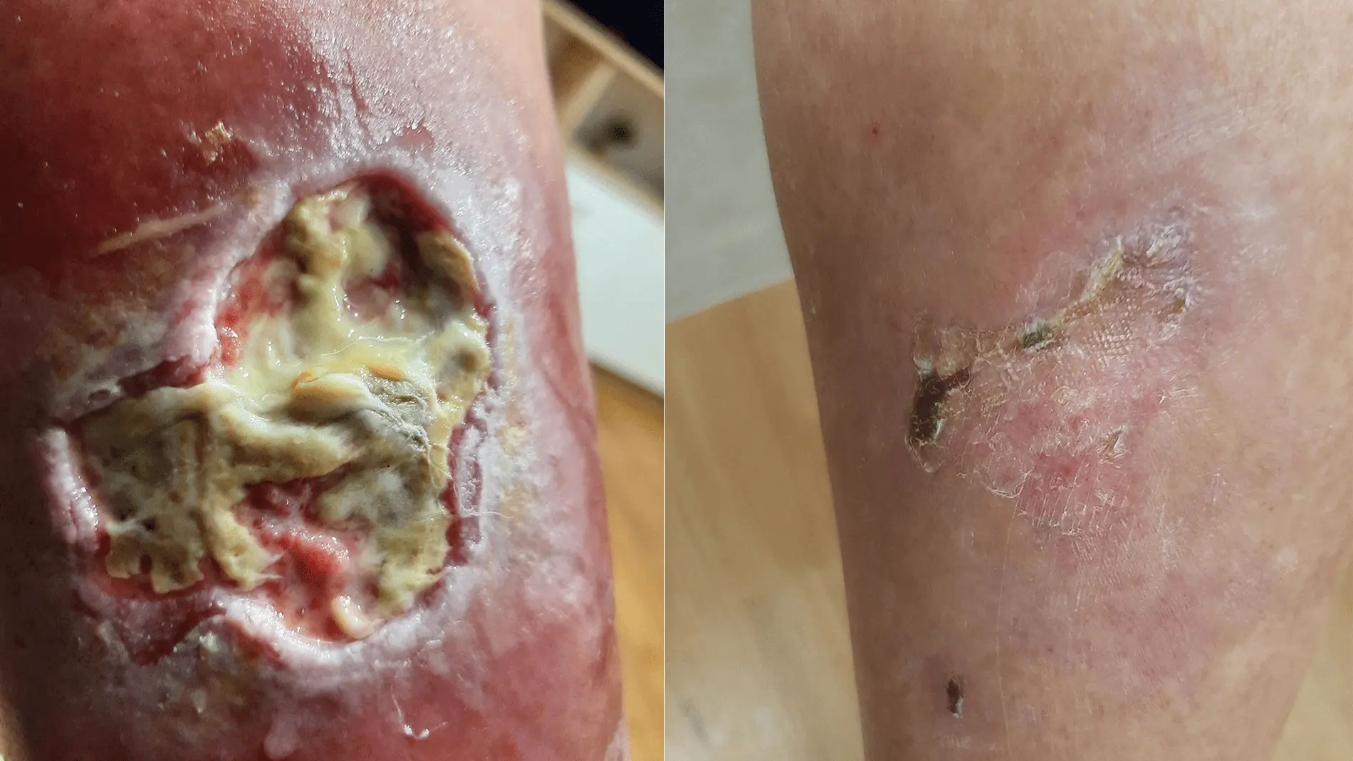
Case in brief
- Patient
- Male, 64 years old at the start of photographic documentation
- Problem
- Chronic wound on the back of the right lower leg, in the calf region
- Approximate onset
- October 2024, after rubbing from a boot while mushroom picking
- Important associated factors
- Type 2 diabetes, treated with oral medication; no insulin
- Previous care
- Several months of visiting-nurse care with various medical preparations; the specific products are not known
- Start of Smrekovit Klasik use
- Early February 2025; the photographs from 3 February mark the time point of initiation
- Photographic course before use
- 11 preserved photographs from 31 December 2024 to 29 January 2025 without a clear trend towards wound closure
- Final documented state
- 5 April 2025 – the area appeared largely closed, with minor residual superficial changes
- Documentation
- 20 original photographs with preserved time stamps and a retrospective in-person interview with the user
Smrekovit Klasik was used as part of the wound care. The case documents a marked temporal change in the course after use began, but as an individual retrospective observational case it does not prove the causal effectiveness of a single intervention.
Table of contents
Baseline condition and previous care
The wound developed on the back of the right lower leg, in the calf region. According to the user, a boot rubbed against this area while he was mushroom picking and caused an injury to the skin. Based on the reconstructed timeline, this most likely occurred around October 2024.
The user had type 2 diabetes, treated with oral medication rather than insulin. Information on long-term blood glucose control, HbA1c values and an objective assessment of circulation in the leg was not available for this case.
During the following months, the wound was cared for by a visiting nurse. The user recalled that various medical preparations had been used, but he could no longer identify the specific products when the case was documented retrospectively. He described the course before Smrekovit Klasik was introduced as the wound becoming “only worse”.
The first preserved photograph is dated 31 December 2024. Eleven photographs are available up to the end of January 2025. They show that the wound remained open, with surface coverings and varying degrees of redness in the surrounding skin, without a clear photographic trend towards closure.
Use of Smrekovit Klasik and wound monitoring
After the wound had persisted for several months, the user began using Smrekovit Klasik in early February 2025. During a later joint review of the photographic series, he identified the photographs from 3 February 2025 as the time when use began. It cannot be established whether they were taken immediately before the first application or shortly afterwards, so this date is treated as the time point marking the start of use.
At the start, he received the usual instructions for using Smrekovit Klasik on an open wound. The user stated that he followed these instructions. However, the exact dressing routine actually used – including the type of gauze, amount of ointment and frequency of dressing changes – was not specifically documented during the course of the case.
The visiting nurse continued to monitor and care for the wound after Smrekovit Klasik was introduced. According to the user, no other new treatment or major change in wound care was introduced at the same time that could be linked to the same temporal turning point.
After the initial advice, the case was not actively followed remotely by Smrekovit. The subsequent course is therefore documented primarily through the original photographs taken by the user himself and his later account of the course.
Photographic course of the wound
The wound was already clearly open in the first preserved photograph
The first preserved photograph was taken on 31 December 2024. According to the reconstructed timeline, the injury had probably already been present for around two months, as the user associated its onset with the mushroom-picking season around October 2024.
The photograph shows an open, irregularly shaped wound with an extensive pale-yellow covering and marked redness of the surrounding skin. A smaller, separate skin lesion is also visible just below the main wound.
This photograph marks the beginning of the preserved photographic documentation, not the onset of the wound itself. According to the reconstructed timeline, Smrekovit Klasik had not yet been introduced at this stage.
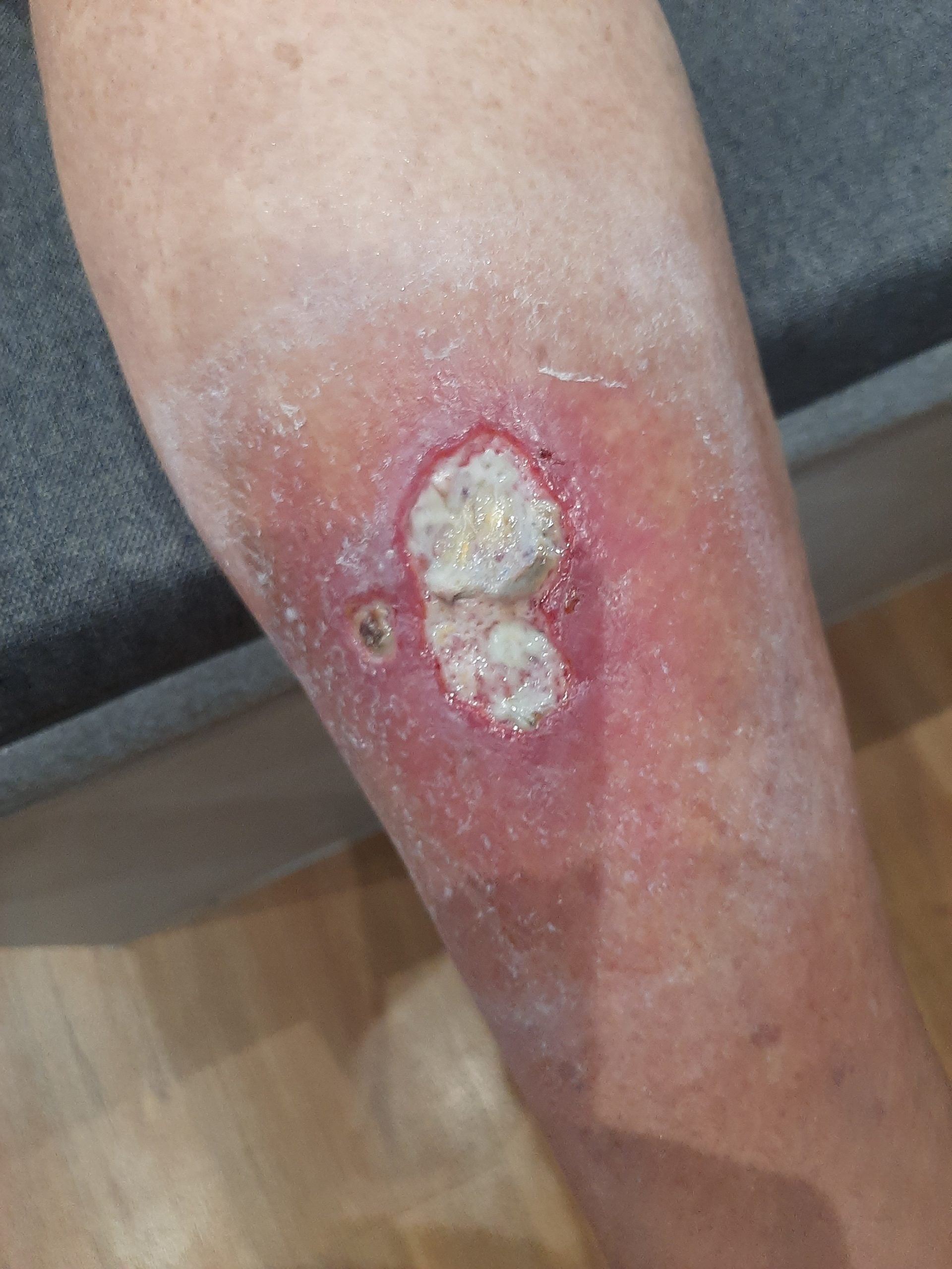
31 December 2024 – first preserved photograph. According to the reconstructed timeline, the wound had probably already been present for around two months; Smrekovit Klasik had not yet been introduced.
Nearly three weeks later, there is still no clear trend towards wound closure
In the photograph from 18 January, the wound remains clearly open and irregular in shape. Yellowish surface coverings are present, and the surrounding skin remains markedly red.
Compared with the first preserved photograph from 31 December, there is no convincing visible progression towards closure. This is one of several consecutive January documentation points showing persistence of the wound before Smrekovit Klasik was introduced.
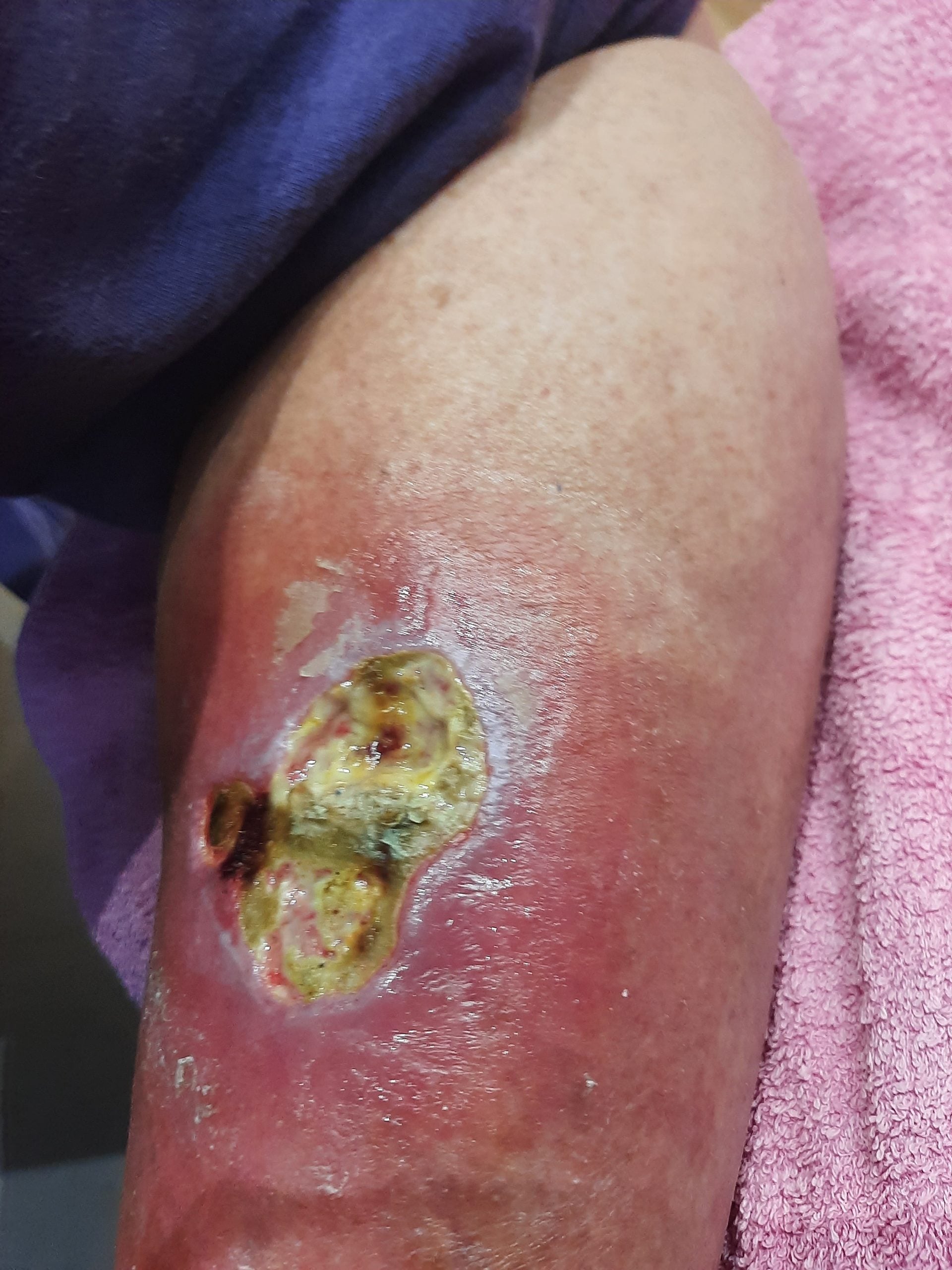
18 January 2025 – the wound remains clearly open. Compared with the first preserved photograph, there is no clear visible trend towards closure.
By the end of January, the wound remained clearly open
In the close-up photograph from 29 January, the wound is still clearly open. Extensive yellow-brown surface coverings, irregular margins and redness of the surrounding skin are visible, together with several darker areas.
This photograph is particularly important because it was taken only a few days before the time point that the user identified, during a joint review of the series, as the start of Smrekovit Klasik use. Even in this last selected January photograph, there is still no clear visible trend towards closure.
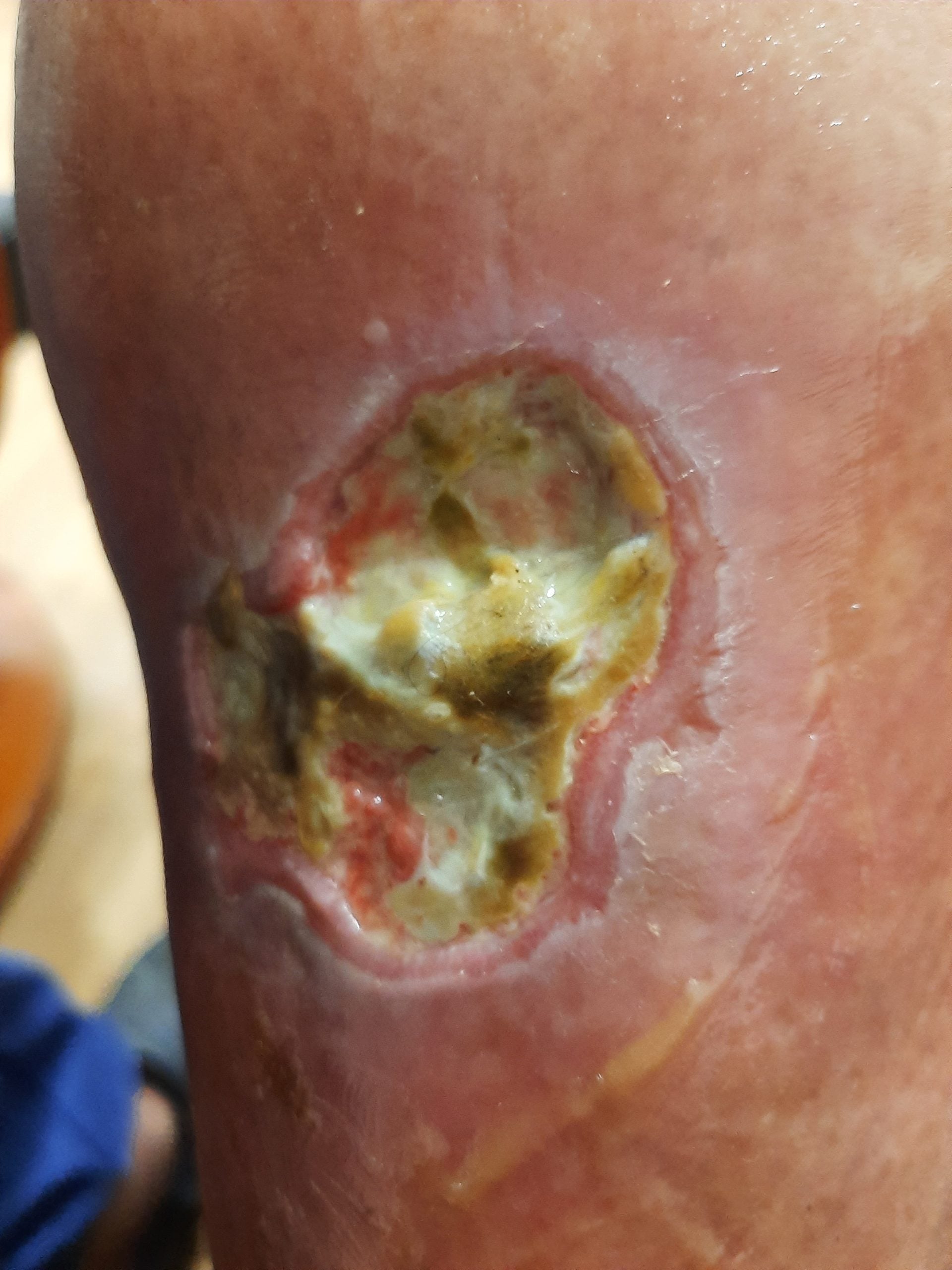
29 January 2025 – the wound remains clearly open only a few days before the time point marking the start of Smrekovit Klasik use.
At the start of use, the wound was still extensively open
Two photographs were taken on the morning of 3 February, approximately one minute apart. In both, the wound remains widely open, with an extensive pale-yellow covering, individual red areas and marked redness of the surrounding skin.
During a joint review of the photographic series, the user identified this date as approximately when he began using Smrekovit Klasik. The preserved documentation does not establish whether the photographs were taken immediately before the first application or shortly afterwards.
For this reason, 3 February is not treated as a strictly defined “before use” image, but as the baseline time point for assessing the subsequent course.
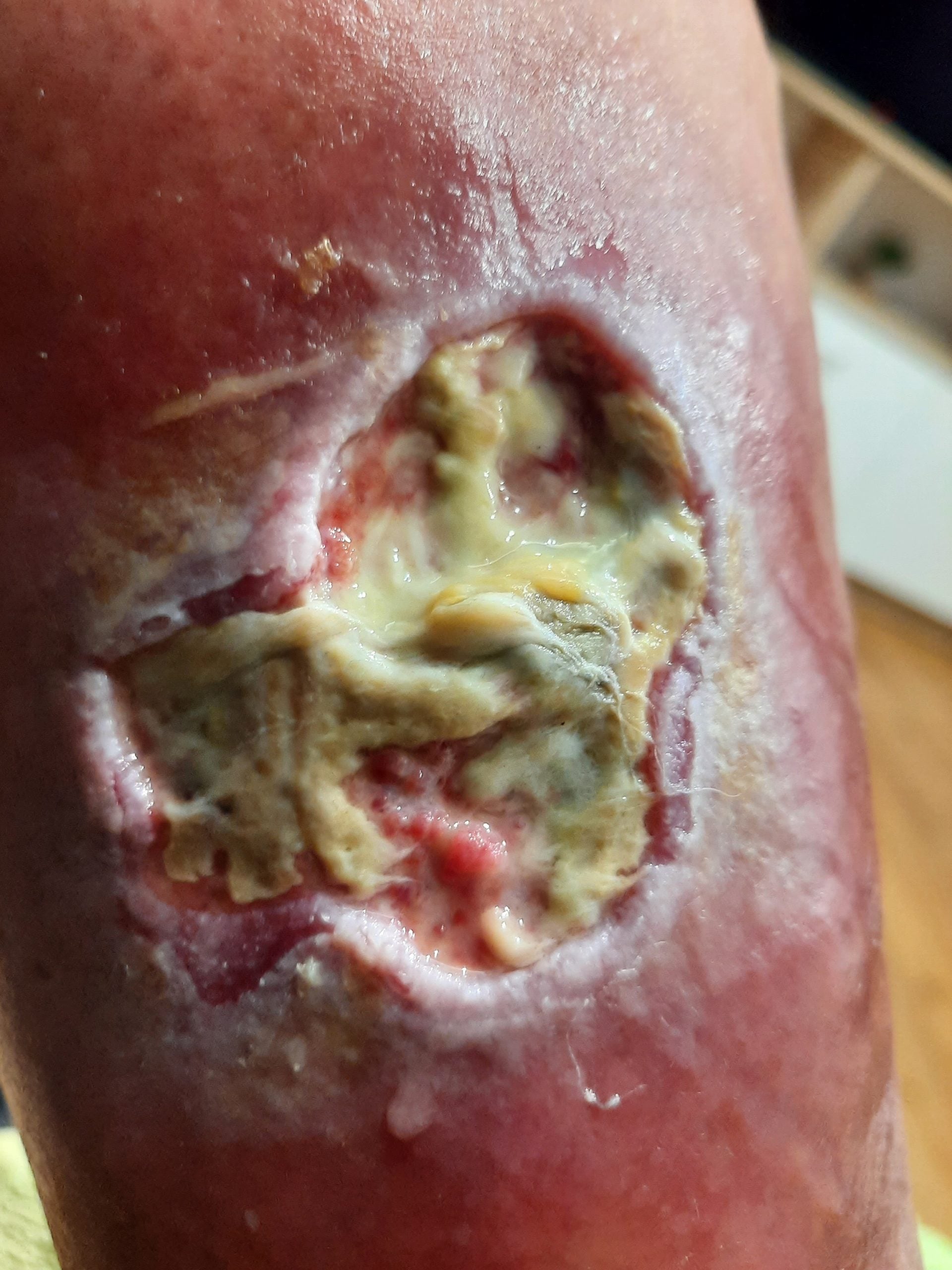
3 February 2025 – during a joint review of the photographic series, the user identified this date as approximately when Smrekovit Klasik use began. It cannot be established whether the photograph was taken immediately before the first application or shortly afterwards.
After approximately three weeks, a marked change in the appearance of the wound is visible
In the photograph from 23 February, a marked change is visible compared with the image from the beginning of the month. There is substantially less pale-yellow surface covering, a larger area of pink-red tissue is visible, and the open area appears smaller and shallower.
Redness of the surrounding skin also appears less pronounced. This is the first photograph in the series after the start of Smrekovit Klasik use to show a clear photographic shift compared with the January images and the state documented on 3 February.
The photograph documents a temporal change in the course of the wound. On its own, it does not establish that Smrekovit Klasik was the direct or sole cause of the observed improvement.
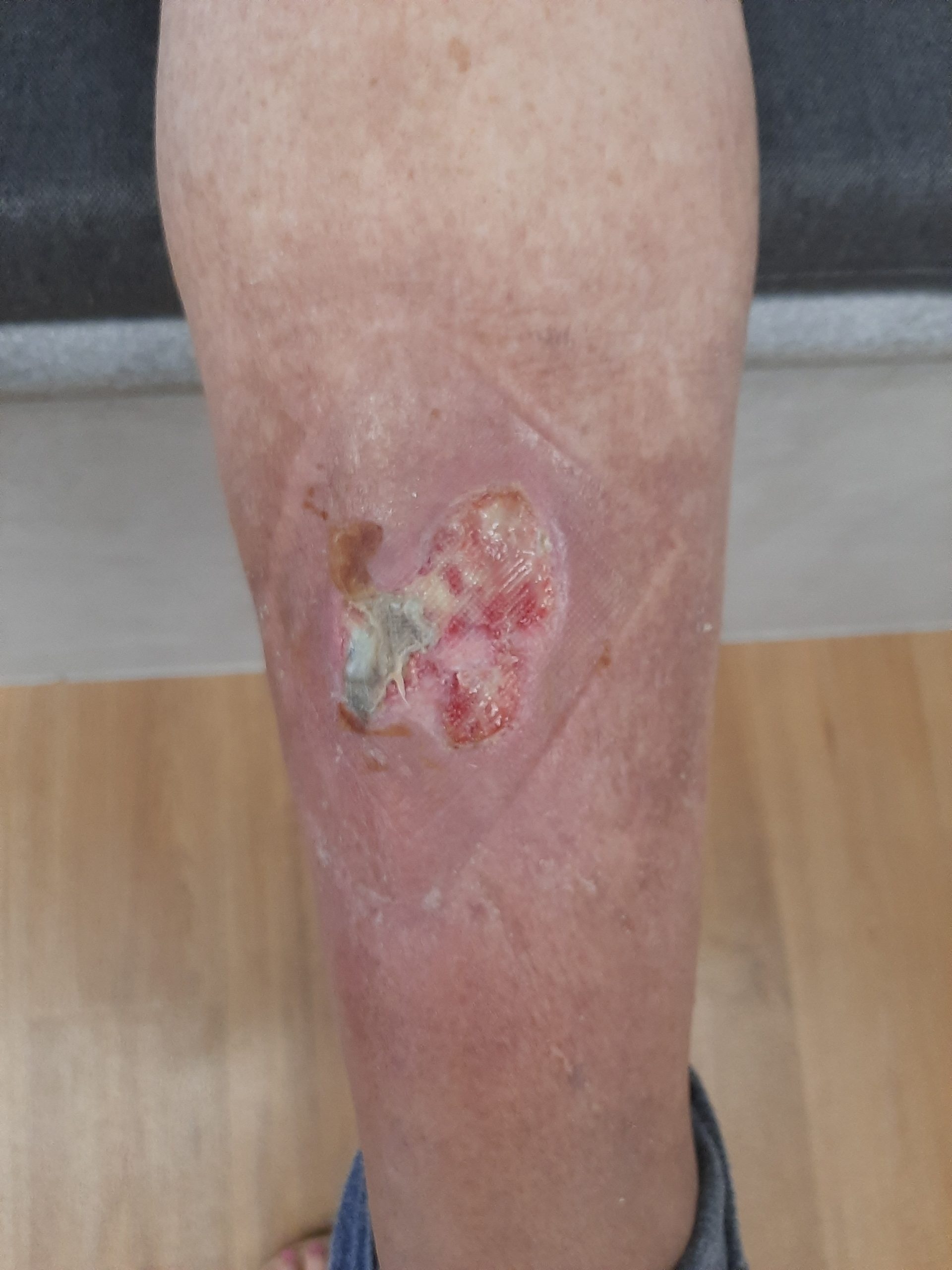
23 February 2025 – approximately 20 days after the time point marking the start of use. Compared with 3 February, there is less pale surface covering, more visible pink-red tissue and a smaller open area.
Five days later, the trend towards a smaller open area continues
In the photograph from 28 February, the open area is smaller than in the earlier February images. Most of the visible surface is pink-red, while the pale-yellow covering is largely confined to a smaller area in the lower part of the wound.
This photograph is particularly useful because it supports the interpretation that the change seen on 23 February was not merely the result of a single image, angle or temporary appearance of the wound, but part of a continuing visual trend over the following days.
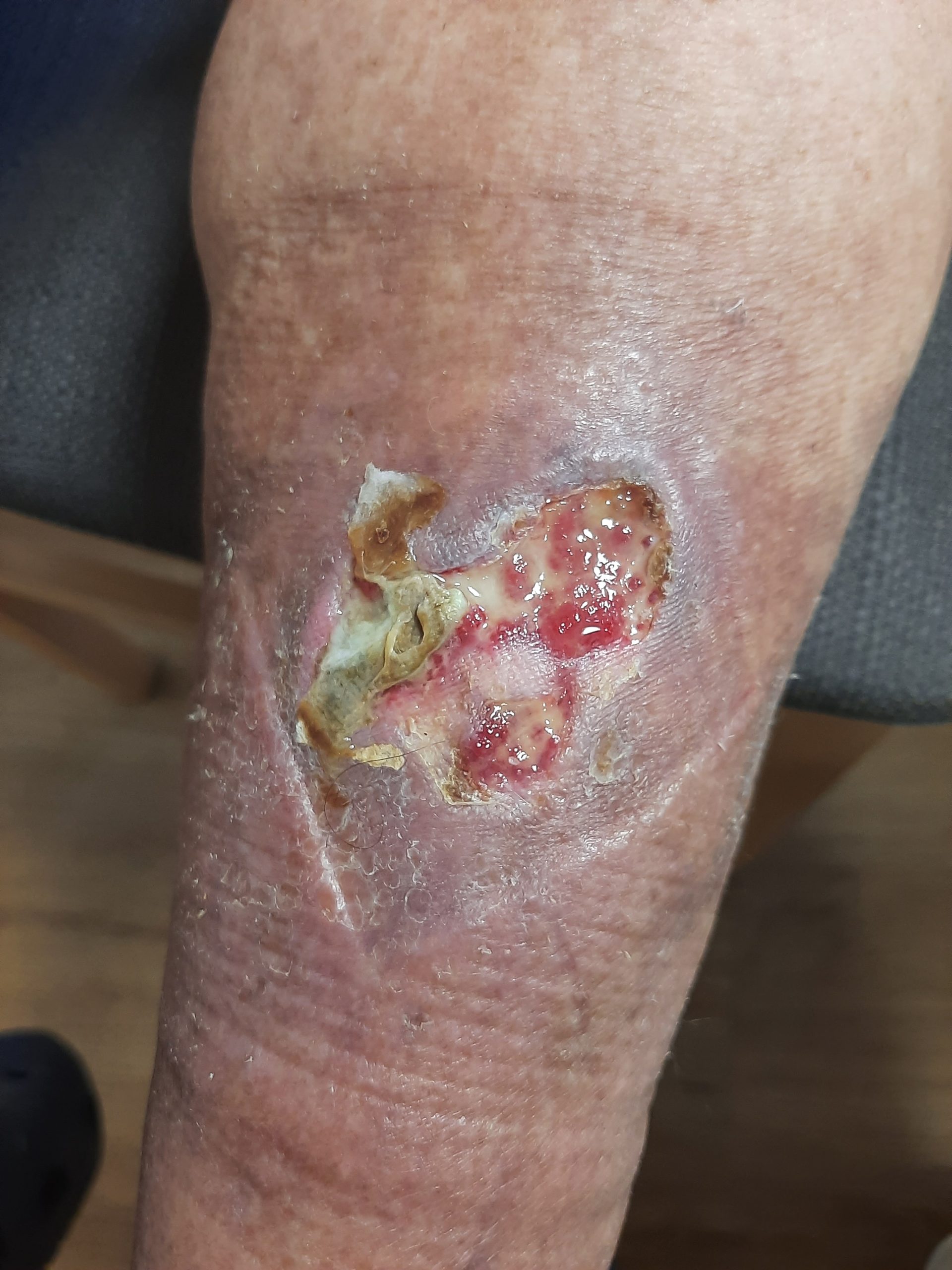
28 February 2025 – the open area is smaller than on 23 February. Pink-red tissue predominates, while the pale covering remains limited to a smaller area.
After approximately six weeks, only a small superficial open area remains
In the photograph from 20 March, the affected area is substantially smaller than at the beginning of February. Only a small elongated superficial open area with a pink-red base remains.
The photographic appearance is consistent with the user’s later estimate that, after starting Smrekovit Klasik, the wound was largely healed within approximately six weeks.
As the photographs were not taken with a measuring scale or under standardised conditions, the exact reduction in wound area cannot be calculated. However, the visual difference compared with the February images is marked.
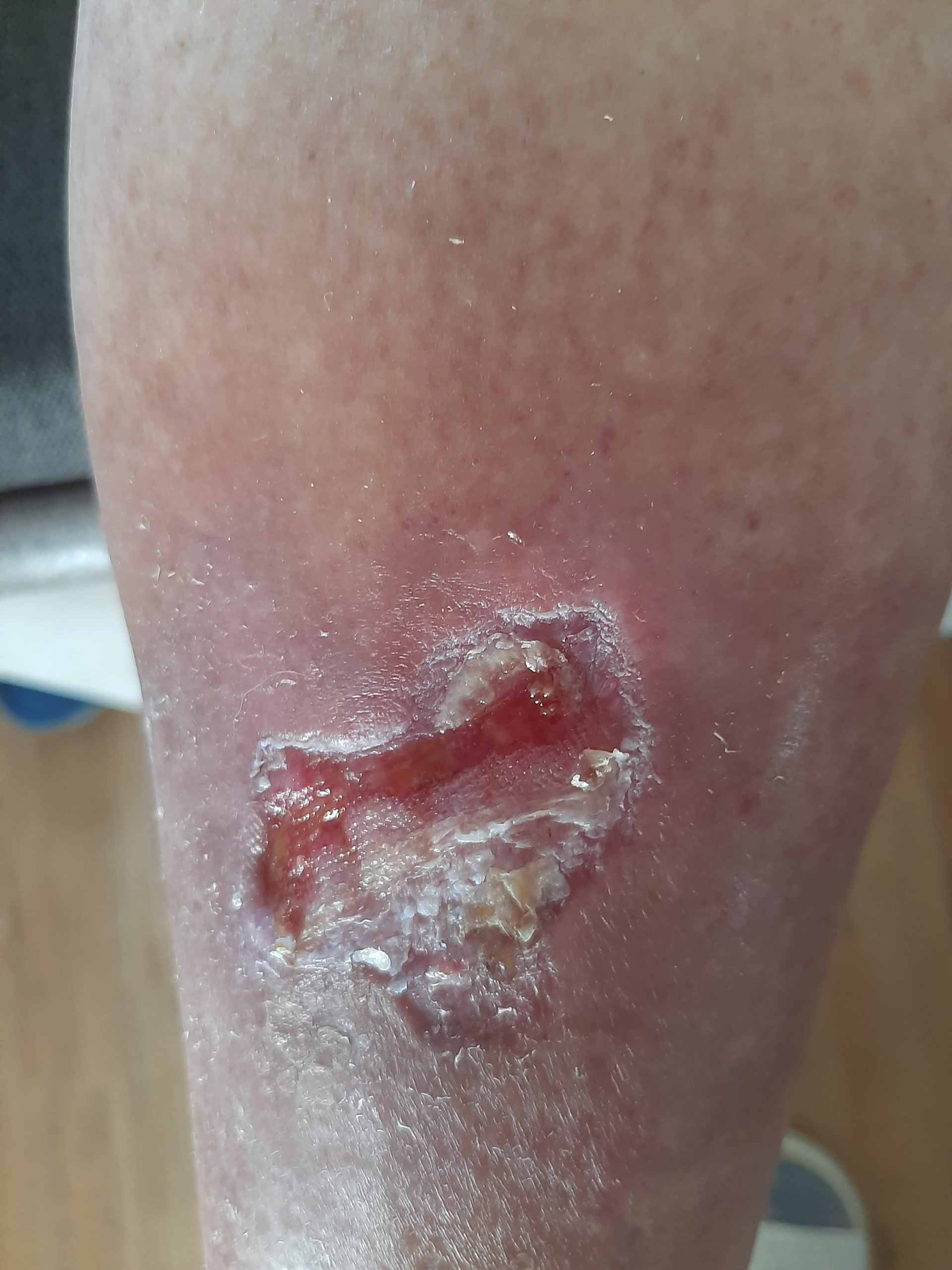
20 March 2025 – approximately 45 days after the time point marking the start of use. Only a small superficial open area remains compared with the extensively open wound documented at the beginning.
By the end of follow-up, the area was largely closed
The final preserved photographs were taken on 5 April 2025. In the sharpest final image, the area of the former wound appears largely closed. A small crusted area remains, while the surrounding skin is dry and mildly red.
Compared with the beginning of February, the open area has virtually disappeared. The final photograph therefore completes a continuous photographic course from a persistent open wound at the end of 2024 to near-complete closure by early April 2025.
As no later photographs are available, 5 April represents the final documented photographic time point in this case.
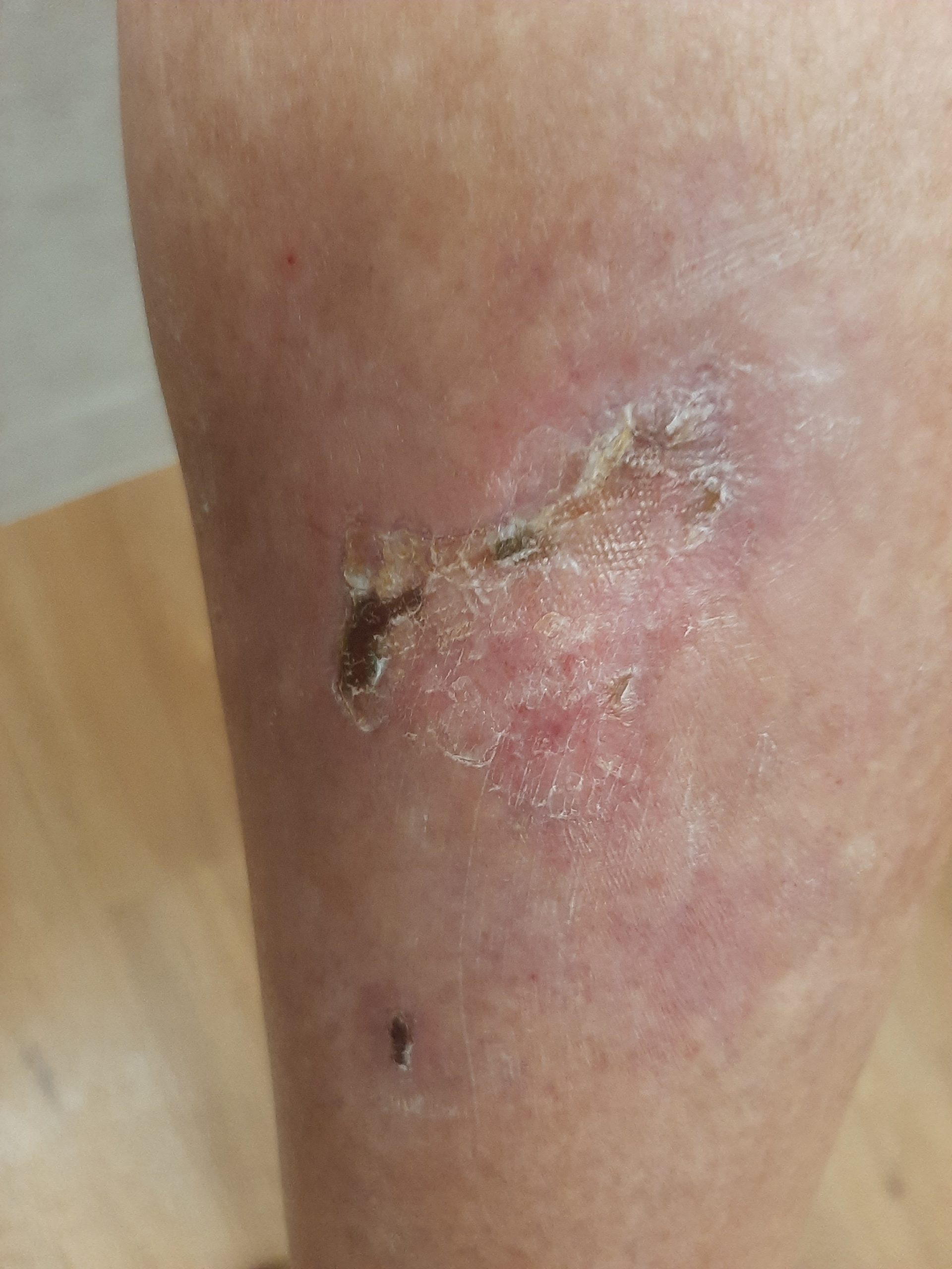
5 April 2025 – final preserved photograph. The area of the former wound is largely closed, with a small residual crusted area and mild surrounding redness.
What the case shows and what it does not show
The strongest aspect of the documentation in this case is not a single “before and after” photograph, but a sequential photographic series before and after the time point marking the start of Smrekovit Klasik use.
Eleven photographs are preserved from the period before use, between 31 December 2024 and 29 January 2025. They show the wound remaining clearly open, without a convincing photographic trend towards closure. On 3 February, at the time point marking the start of use, the wound was still extensively open.
In the photograph from 23 February, approximately 20 days later, a marked change is visible for the first time: less surface covering, more visible pink-red tissue, and a smaller open area. Subsequent photographs from 28 February, 20 March and 5 April show a continuous trend towards a smaller open area and eventual near-complete closure.
During the retrospective interview, the user stated that no other new treatment or major change in wound care was introduced when Smrekovit Klasik was started. Visiting-nurse care did, however, continue, so Smrekovit Klasik was not the only factor present during the period in which the wound closed.
The temporal sequence is therefore notable and well documented, but an individual observational case cannot prove that Smrekovit Klasik was the direct or sole cause of wound closure. The outcome also cannot automatically be generalised to other people or other types of wounds.
Documentation and limitations of the case
The case is documented by 20 original photographs taken by the user himself. The photographs contain preserved time stamps from 31 December 2024 to 5 April 2025 and form a chronologically continuous series.
Information about the onset of the wound, previous care, the start of Smrekovit Klasik use and relevant associated factors was additionally collected during a retrospective in-person interview with the user on 7 October 2026.
A discrepancy in the years became apparent in the oral account: the user initially placed the onset of the wound in October 2025 and the start of Smrekovit Klasik use in February 2026. This is inconsistent with the original photographic series. Because the photographic time stamps, their chronological sequence and the user’s identification of the start of use during the joint review are mutually consistent, the photographic timeline was used for reconstruction of the case: onset around October 2024 and start of use in early February 2025.
The photographs were not taken under standardised conditions: angle, distance and lighting vary, and no measuring scale is present. Therefore, wound area or depth and the exact rate of reduction cannot be reliably calculated from the photographs.
No laboratory data on diabetes control, such as HbA1c, were obtained for this case, nor was there an objective assessment of arterial or venous circulation in the lower limb. Independent medical records or visiting-nurse documentation were also not obtained.
The specific medical preparations used before Smrekovit Klasik was introduced are unknown. The exact dressing technique, amount of ointment used and frequency of dressing changes were likewise not documented prospectively.
The report was prepared by Smrekovit on the basis of material supplied by the user. As the report’s preparer is associated with the manufacturer of Smrekovit Klasik, this represents a potential conflict of interest and a possible source of interpretative bias.