
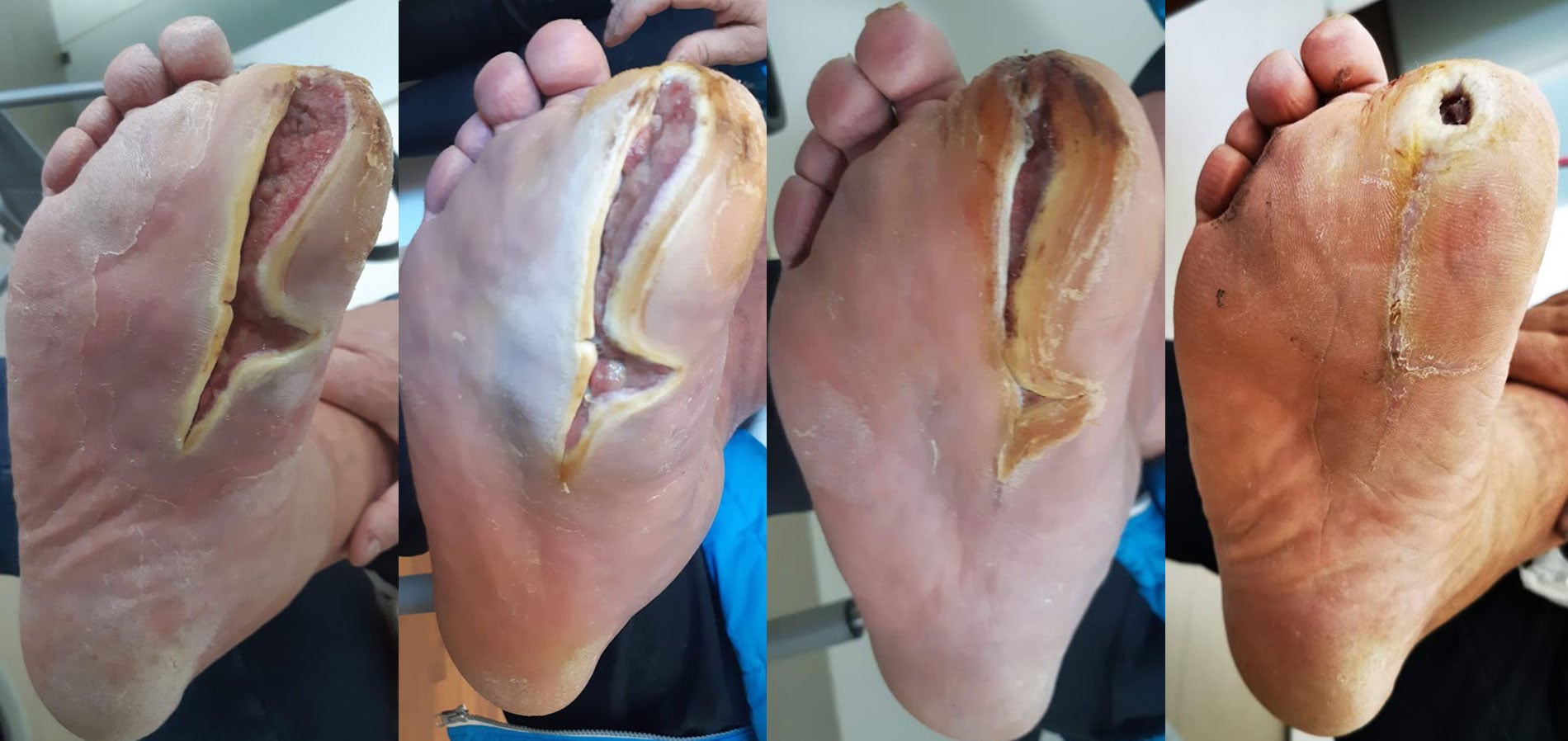
Case in brief
- Patient
- Middle-aged man; approximately 50–60 years old by retrospective estimate
- Underlying condition
- Diabetes and diabetic foot
- Previous course
- Several debridements; the last more extensive procedure took place several months before follow-up began
- Start of documented use
- Initial open wound area
- 16.47 cm²
- Final measurement
- 1.16 cm² after 155 days
- Reduction in open wound area
- Approximately 93%
- Use
- Smrekovit Klasik on sterile gauze, generally once daily
- Documentation
- A report prepared in 2020, four dated photographic checkpoints, measurements of the open wound area and later clarifications by the author
At the last documented follow-up the wound was not yet completely closed. The case has no control group or preserved independent medical records, so it cannot establish a causal effect of any single intervention.
Table of contents
Baseline: chronic diabetic foot wound
The patient was a middle-aged man with diabetes and a history of diabetic foot complications. Several years earlier, a toe on the right foot had been amputated because of a previous diabetic foot complication. The amputation wound had completely healed before the present case and was not part of the wound followed here.
The current wound on the right foot had not closed satisfactorily before follow-up began. It had undergone several surgical debridements. According to the report prepared in 2020, the last and relatively extensive debridement had been performed several months before the first documented assessment. The wound remained open afterwards.
At the start of documented follow-up on 15 January 2020, the measured open wound area was 16.47 cm². The wound was surrounded by callused skin, with a clearly defined boundary between the open area and the surrounding skin.
According to the author’s later recollection, the patient had markedly reduced sensation in the foot, consistent with diabetic neuropathy, and therefore barely felt the wound and did not report pain. This information is not supported by preserved neurological records and is therefore treated as retrospective information.
How was the wound treated?
Documented use of Smrekovit Klasik began on 15 January 2020. Under the protocol used at that time, an approximately 1–2 mm layer of ointment was applied to sterile gauze. The gauze was then placed directly on the wound with the ointment-coated side facing the wound and secured with medical tape.
Directly supervised wound care is documented on 15 January, 22 January and 27 February 2020. The patient was instructed to repeat the same procedure at home once daily. At the follow-up visits, the wound was photographed before applying a new dressing.
The original protocol also included gentle removal of any remaining ointment using a sterile stainless-steel spatula. This detail is retained for an accurate historical reconstruction of the case and does not represent the current recommended procedure.
According to the author’s later recollection, the patient continued daily local use of Smrekovit Klasik throughout the documented period, with no further debridements or other local preparations during that time. This information is based on retrospective recollection and is not supported by independent medical records.
The current method of use for open wounds is described separately in the wound care instructions.
Measurement of the open wound area
The open wound area was quantitatively measured from photographs containing a measurement scale at four documented follow-up points. The photographs were graphically adjusted to a common scale and the open area was then measured using the measurement tool in Adobe Acrobat Pro.
The original 2020 report already stated an estimated measurement uncertainty of ±15%. The measurements were not recalculated during the retrospective update; the originally documented values were retained.
| Date | Day of follow-up | Open wound area | Reduction from baseline |
|---|---|---|---|
| 15 Jan 2020 | 0 | 16.47 cm² | 0% |
| 22 Jan 2020 | 7 | 14.90 cm² | 9.5% |
| 27 Feb 2020 | 43 | 5.29 cm² | 67.9% |
| 18 Jun 2020 | 155 | 1.16 cm² | 93.0% |
The reduction from 16.47 to 14.90 cm² during the first seven days was approximately 9.5%, which is smaller than the stated ±15% measurement uncertainty. This early change should therefore not be interpreted on its own as a reliable quantitative reduction.
At the later follow-ups the differences were substantially larger. After 43 days, the measured open wound area was approximately 67.9% smaller than at baseline, and after 155 days it was approximately 93.0% smaller. Only two-dimensional wound area was measured; wound depth and volume were not quantified.
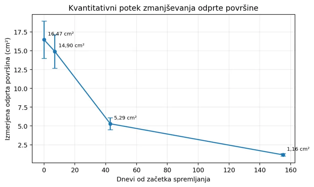
Measured open wound area during documented follow-up. Vertical bars represent the estimated ±15% measurement uncertainty stated in the 2020 report.
Photographic course of the wound
Open wound area 16.47 cm²
At the first documented assessment, a relatively large open wound was present on the right foot, surrounded by pronounced callused skin. The boundary between the open wound and the surrounding skin was clearly defined.
The measured open wound area was 16.47 cm². The original report did not describe visible signs of necrosis at this assessment. This measurement serves as the baseline for all subsequent comparisons.
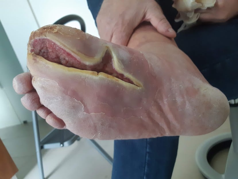
15 January 2020 – initial documented state; measured open wound area 16.47 cm².
First follow-up: 14.90 cm²
After seven days, the measured open wound area was 14.90 cm². The original report describes less pronounced redness in the central part of the wound and formation of new epithelium along the boundary between the wound and the callused skin.
The measured area was approximately 9.5% smaller than at baseline. Because the estimated measurement uncertainty was ±15%, this early difference should not on its own be regarded as a reliable quantitative reduction.
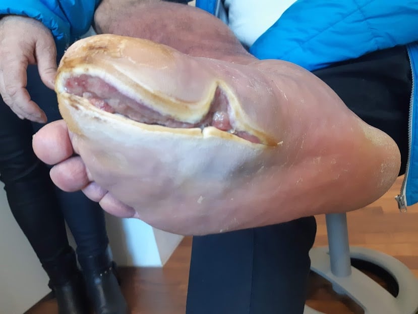
22 January 2020 – first follow-up after seven days; measured open wound area 14.90 cm².
Open wound area reduced to 5.29 cm²
At the third documented follow-up, the open wound area was markedly smaller than in January. It measured 5.29 cm².
This represents an approximately 67.9% reduction from the initial 16.47 cm². The difference is substantially larger than the stated measurement uncertainty and represents the first clearly documented quantitative reduction in open wound area in this case.
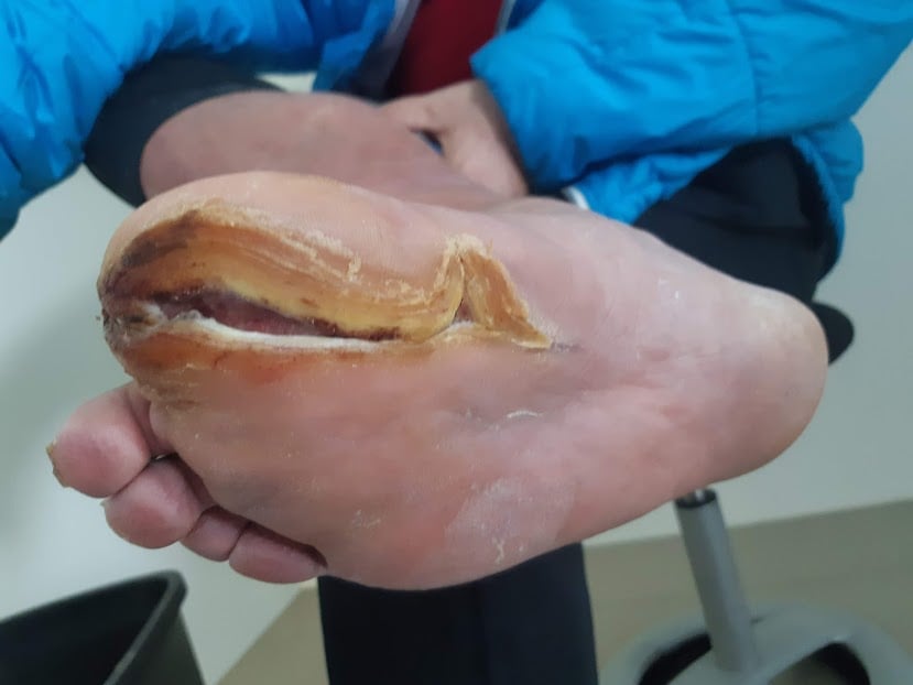
27 February 2020 – open wound area 5.29 cm²; approximately 67.9% smaller than at baseline.
Remaining open wound area 1.16 cm²
On the final preserved follow-up photograph, most of the area that had originally been open appeared closed or epithelialised. A small open area remained.
The measured open wound area was 1.16 cm², representing an approximately 93.0% reduction from baseline. The wound was not yet completely closed at this point, and no further photographic follow-up is preserved after 18 June.
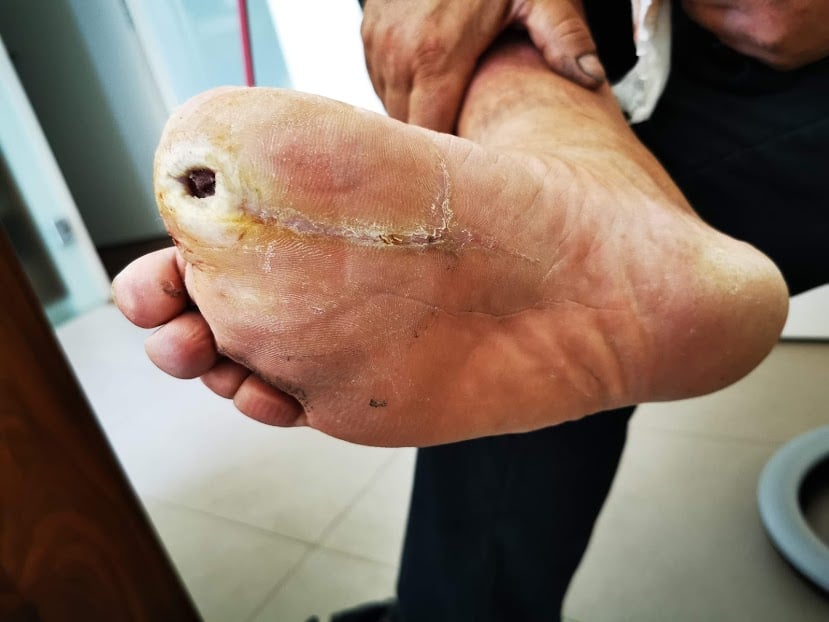
18 June 2020 – final documented follow-up after 155 days; open wound area 1.16 cm², approximately 93% smaller than at baseline. A small open area remains.
What is important about this case?
This case is noteworthy primarily because the wound was not new when documented follow-up began. According to the available information, it had previously undergone several surgical debridements and had remained open for several more months after the last more extensive procedure.
During the following 155 days, progression was documented with dated photographs and wound-area measurements. The measured open wound area decreased from 16.47 cm² to 1.16 cm², representing an approximately 93% reduction. This is more informative than a purely subjective assessment of wound appearance because it allows the change to be followed quantitatively over time.
The case must nevertheless be interpreted cautiously. The patient had diabetes and markedly reduced sensation in the foot, while information on perfusion, diabetes control and other factors affecting wound healing has not been preserved. According to the author’s later recollection, Smrekovit Klasik was the only local preparation used during the observed period and no further debridements were performed, but this cannot be independently verified.
At the final documented follow-up, the wound was not yet completely closed. The case therefore documents a pronounced and measurable reduction in open wound area during a period of regular Smrekovit Klasik use, rather than documented complete healing. A single retrospective case cannot determine how much of the observed course can be attributed to any one intervention.
Documentation and limitations of the case
The case was retrospectively reconstructed from a report prepared in 2020, four dated photographic checkpoints, preserved measurements of the open wound area and later clarifications provided by the author.
An important limitation is that no independent medical records have been preserved regarding the patient’s diabetes, previous debridements, circulation in the foot or other factors that may have affected wound healing. Some information, including the approximate age, markedly reduced sensation in the foot and the use of Smrekovit Klasik as the only local preparation during the observed period, is based on the author’s later recollection.
The photographs were not standardised with regard to angle, distance, lighting or position of the foot. Wound area was determined from two-dimensional photographs, and the original report already stated an estimated measurement uncertainty of ±15%. Wound depth and volume were not measured.
There is a long interval without an intermediate documented measurement between the 27 February follow-up and the final photograph on 18 June. Complete wound closure was also not documented, as a small open area was still present at the final follow-up.
This is a single retrospectively documented case without a control group. The author of the report is affiliated with the manufacturer of Smrekovit products and participated in wound care, measurement and interpretation of the case, which represents a potential conflict of interest and a possible source of interpretative bias.