
Case at a glance
- Patient
- 56 years old at the time of injury
- Date of injury
- Injury
- Crush injury and burn to the left foot caused by a heavy water heater falling on it, with a simultaneous fracture of the distal phalanx of the big toe
- Wound development
- Extensive necrosis developed on the dorsum of the foot; on 16 March, the specialist described it as larger than half a man’s palm
- Early use of Smrekovit Klasik
- According to the patient, already on , the day after the injury
- Documented resumption of systematic use of Smrekovit Klasik
- From , on sterile nonwoven gauze
- Concurrent medical care
- Community nursing dressing changes, other dressings, partial removal of necrotic tissue, necrectomy and Dermazin
- Visual wound closure
- Follow-up photograph
- – the area remains closed with no visible open wound
- Documentation
- Medical reports, community nursing documentation, SMS/MMS correspondence and serial photographic monitoring
Smrekovit Klasik was part of the overall wound care and was not the only intervention used. The case therefore presents the documented course of an individual wound, not the isolated effect of a single intervention.
Table of contents
How did the injury occur?
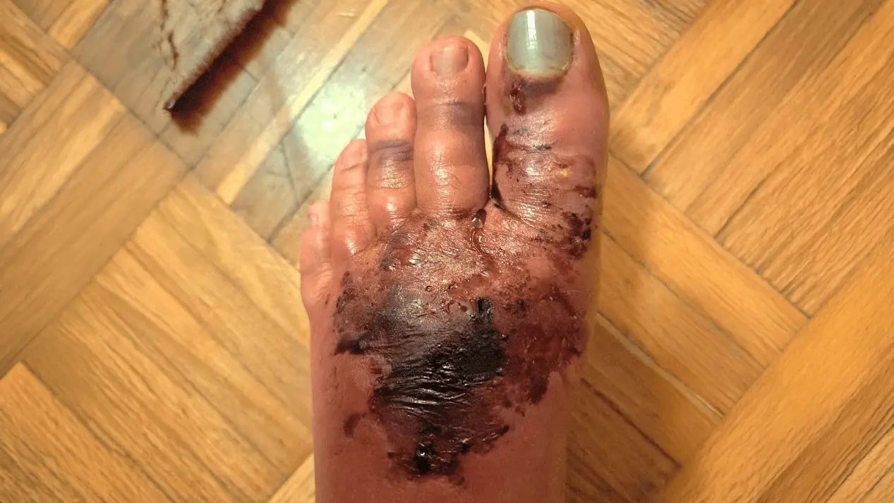
The injury occurred on 12 February 2026, when a heavy water heater fell onto the patient's left foot at home. The incident caused a combined injury to the foot: a severe impact or crush injury together with a hot-water burn.
At the first medical examination, the foot was markedly painful, swollen and bruised, and the skin on the dorsum of the foot was damaged. X-ray imaging also showed a fracture of the distal phalanx of the left big toe. Rest, unloading of the foot, crutches and pain treatment were recommended, and a temporary below-knee splint was also applied.
Over the following days, blisters appeared in the injured area, and later dead or necrotic tissue began to develop on the dorsum of the foot.
According to the patient's own report, she began using Smrekovit Klasik immediately after the injury. A photograph from 13 February 2026, one day after the accident, documents the early condition of the foot during this period of use.
Development of necrosis
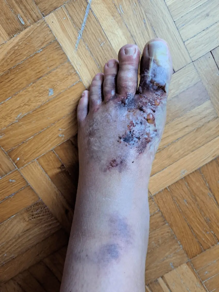
At the follow-up examination on 23 February 2026, eleven days after the injury, an epidermal to superficial dermal injury or burn was described on the dorsum of the foot, together with a small area of dry necrosis. This confirms that tissue necrosis was already present before detailed photographic monitoring began in March.
Over the following days, the condition progressed markedly. In the photograph from 5 March 2026, an extensive, sharply demarcated black necrotic area is already visible on the dorsum of the foot, while the foot remained markedly swollen.
According to the patient’s own report, she had already been using Smrekovit Klasik during the first days after the injury. After she entered medical and community nursing care, the methods of local wound care changed over time. On 4 March 2026, she resumed systematic use of Smrekovit Klasik on sterile nonwoven gauze. Because well-preserved SMS/MMS communication is available from this point onward, this date is used as the beginning of the closely monitored renewed systematic use.
How was the wound treated?
From 4 March 2026, the renewed systematic use of Smrekovit Klasik is documented. The ointment was applied to sterile nonwoven gauze, which was then placed with the coated side directly onto the entire wound. The frequency of dressing changes was adjusted according to the amount of exudate.
Wound care during this period did not follow a single method. The patient was monitored by the community nursing service, and copper or bronze dressings were also used during certain periods. Part of the necrotic tissue was gradually removed.
On 16 March 2026, the wound was examined by a specialist in plastic surgery. The necrotic area was described as larger than half of a man's palm, and necrectomy was performed up to the point of pain. Dermazin was used after the procedure. During the subsequent course, the patient returned to using Smrekovit Klasik on nonwoven gauze.
Because several different procedures were used, this case does not allow the effect of an individual intervention to be separated. It does, however, allow detailed monitoring of the wound course during a period in which Smrekovit Klasik was an important and repeatedly used part of wound care.
Photographic course of the wound
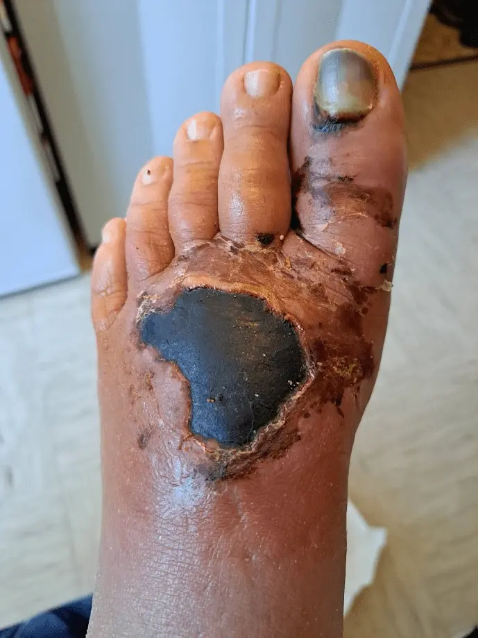
Gradual separation of necrotic tissue
The necrotic eschar was less homogeneous than at the beginning of monitoring. Black-grey, yellowish and isolated reddish areas were visible. During this period, the community nursing service performed dressing changes and partially removed separating necrotic tissue.
Less black necrosis and more open wound surface
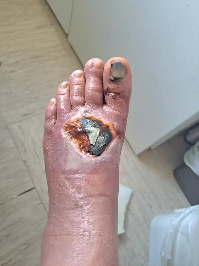
Nine days after the photograph from 13 March, the necrotic eschar was considerably less uniform. The photograph is dominated by yellow-brown areas, isolated black areas of necrosis and already visible reddish areas of the open wound.
In the meantime, on 16 March, the wound was examined by a specialist in plastic surgery, who performed necrectomy up to the point of pain and used Dermazin after the procedure. In the following days, bronze dressings were also used again, and on 22 March the patient returned to using Smrekovit Klasik.
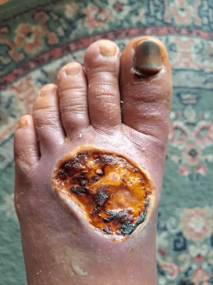
More visible granulation tissue
In the days before the photograph, there was also a problem with one of the dressings: the dressing was left on the wound for too long and adhered as it dried. The patient was therefore again advised to apply Smrekovit Klasik to sterile nonwoven gauze and to change the dressing more regularly.
In the photograph from 27 March, compared with the previous images, more pink-red granulation tissue is visible, while there is less black necrotic tissue. The wound is still extensive and open, but its appearance is clearly changing.
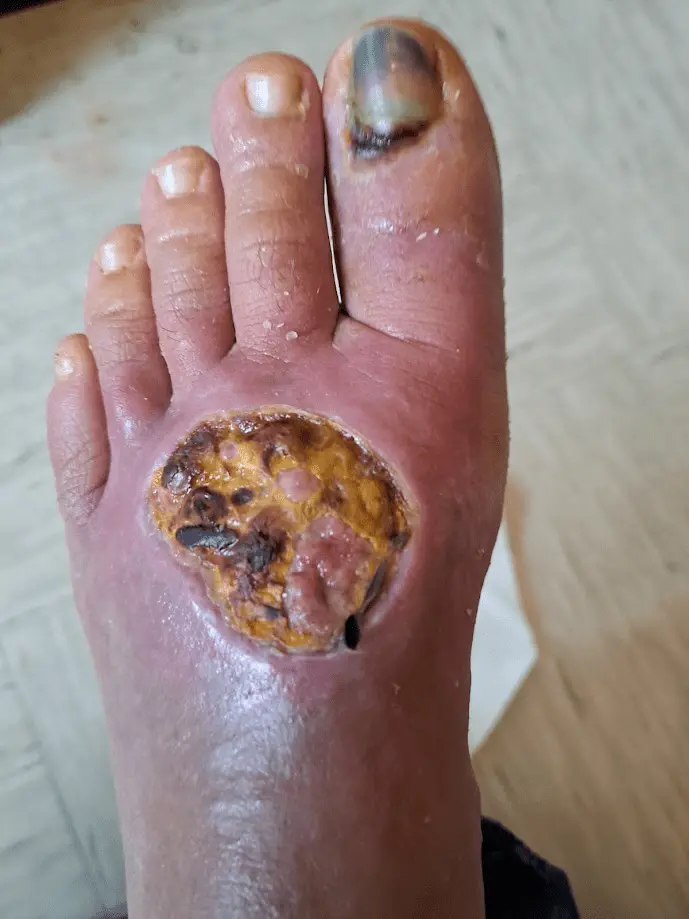
The open wound surface is noticeably decreasing
In the photograph from 13 April, the open wound surface is already markedly smaller compared with the March images. Red granulation tissue predominates, while black necrotic tissue is practically no longer visible.
At this stage, the wound was not yet closed. Exudate and swelling were still present, and the patient continued to report problems related to the foot injury itself.
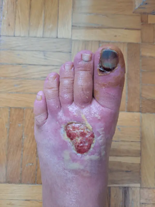
Further reduction of the wound
Four days before this photograph, the wound was again examined by a specialist in plastic surgery. In the report dated 16 April 2026, the specialist noted that the wound had decreased nicely in size and that an unhealed area of approximately 3–4 cm remained. Hypergranulation tissue was also present.
In the photograph from 20 April, the open wound surface is even smaller than one week earlier. Granulation and gradual wound closure are continuing. At this stage, the tissue was not yet fully mechanically stable, as the patient reported that the wound could still bleed with greater loading or while driving.
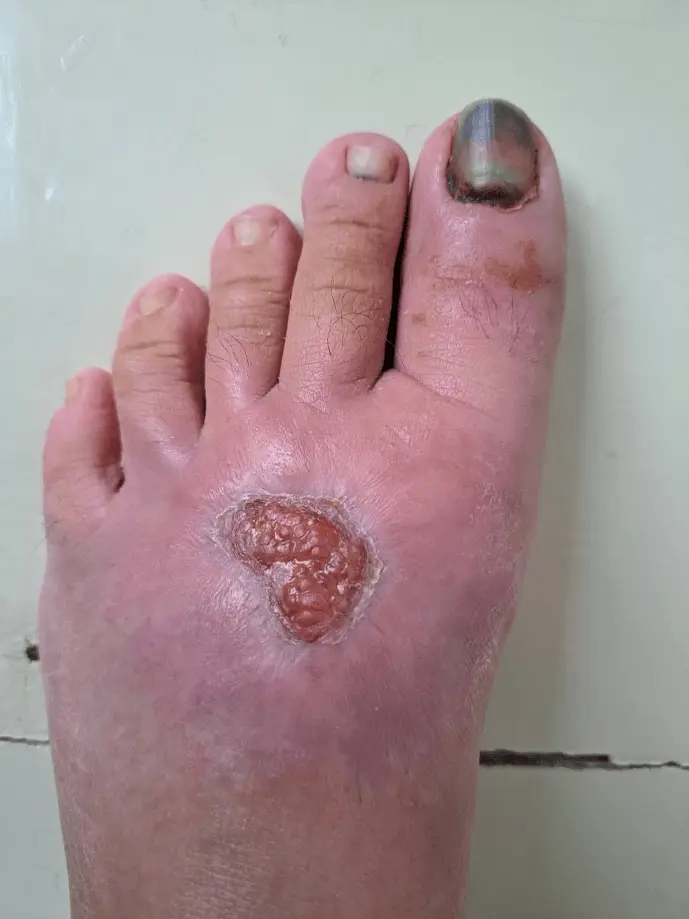
The wound is visually closed
In the photograph from 2 May, no open wound surface is visible anymore. The area is practically epithelialised or closed, although a small superficial skin change remains.
In the time-stamped communication, the wound was also described that same day as nicely closed. The community nursing service nevertheless continued to document dressing changes until 13 May 2026.
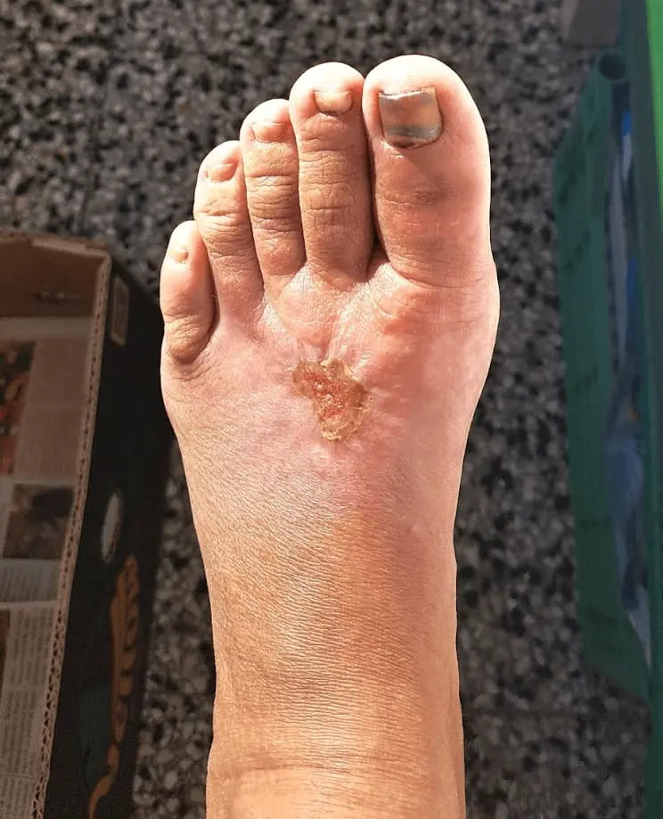
Follow-up photograph: the wound remains closed
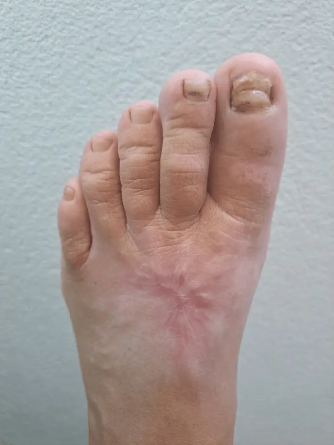
The follow-up photograph from 6 June 2026, approximately one month after the visual closure of the wound, shows a completely closed area with no visible open wound.
A relatively calm change in skin colour and texture remains at the site of the injury, but there is no visible reopening of the wound in the photograph. This concludes the photographic monitoring of the skin aspect of the case.
What is important about this case?
The case is particularly notable because of the extent of the initial injury and the well-documented course. Over several weeks, the large black necrotic area gradually changed into an open wound with granulation tissue, after which the wound surface continued to decrease until complete closure.
An important feature is that the necrosis in this case did not develop because of a known chronic vascular disease or diabetes, but following a severe mechanical injury and burn to the foot. According to information provided by the patient during follow-up, she was not aware of having diabetes, venous insufficiency, heart failure or another major chronic condition that would markedly impair wound healing.
At the same time, the course was not idealised. During recovery, the foot was repeatedly subjected to considerable strain from walking on stairs, driving and everyday obligations. After greater exertion, the patient also reported increased pain, swelling or renewed bleeding.
Smrekovit Klasik was used regularly on sterile nonwoven gauze during a large part of the documented period, but the wound was also receiving community nursing and specialist care at the same time. The case therefore does not allow the precise effect of each individual procedure to be separated, but it provides a very clear picture of the actual multi-week course of a complex post-traumatic wound.
Documentation and limitations of the case
The case was reconstructed retrospectively from several complementary sources: medical reports, community nursing documentation, time-stamped SMS/MMS communication and serial photographic monitoring.
The medical documentation includes treatment at the time of injury, a trauma follow-up, two examinations by a specialist in plastic and reconstructive surgery, and later imaging examinations. The community nursing service documented dressing changes between 16 February and 13 May 2026.
When interpreting the case, it is important to note that wound care was not standardised. In addition to Smrekovit Klasik, other dressings, Dermazin and surgical removal of necrotic tissue were also used at different times. There was also no control group or standardised measurement of wound surface area.
This is therefore a single documented observational case showing the actual course of the wound and the use of Smrekovit Klasik as part of the overall wound care. It cannot be used to directly predict the course or outcome of other wounds.