
Case at a glance
- Patient
- 77 years old at the time of injury
- Date of injury
- Injury
- Post-traumatic wound of the lower leg after striking it against a motorcycle starter
- Factors affecting healing
- Venous and heart failure; the severity of both conditions is not objectively defined in the preserved documentation
- Initial open wound area
- 25.08 cm² at the beginning of documented monitoring on 4 August 2018
- Documented use of Smrekovit Klasik
- From to , on sterile gauze
- Measured course
- 25.08 cm² → 15.43 cm² after 5 days → 0.20 cm² after 12 days → 0 cm² after 19 days
- Wound closure
- – both areas were considered epithelialised at the time
- Documentation
- Original report from 2018, brief questionnaire, serial dated photographs, measurement appendices and wound-area calculations
The case is particularly notable for its serial photographs and quantitative measurement of the open wound area. Measurements were made from photographs with a measurement scale, with a measurement uncertainty of ±15% already estimated in the original report. This is a single documented observational case, so the course cannot establish the causal effectiveness of any individual intervention.
Table of contents
How did the injury occur?
The injury occurred on 19 July 2018, when the 77-year-old patient struck the lower part of his leg against a motorcycle starter. The impact caused a localised post-traumatic wound, which was later designated as wound 1 in the original documentation.
Because the condition did not improve satisfactorily after the injury, according to the preserved questionnaire the patient visited a doctor approximately three days later and then attended regular dressing changes for about two weeks.
In the period before documented monitoring began, a substantially larger superficially damaged area developed above the original wound, designated as wound 2. The original 2018 report associated its development with the use of a polyurethane dressing with a waterproof film, but the original medical documentation from that period has not been preserved, so this causal relationship cannot now be confirmed reliably.
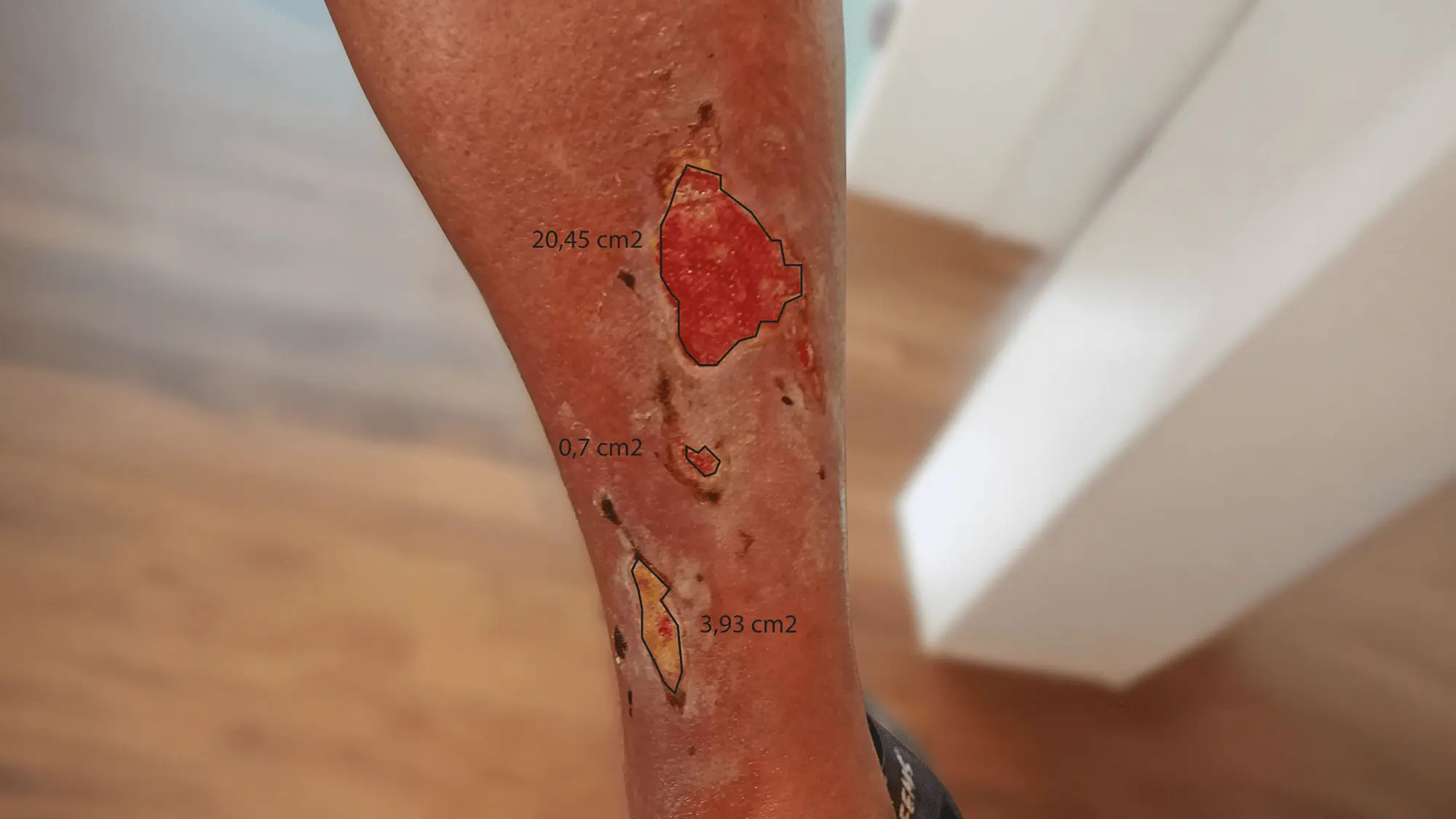
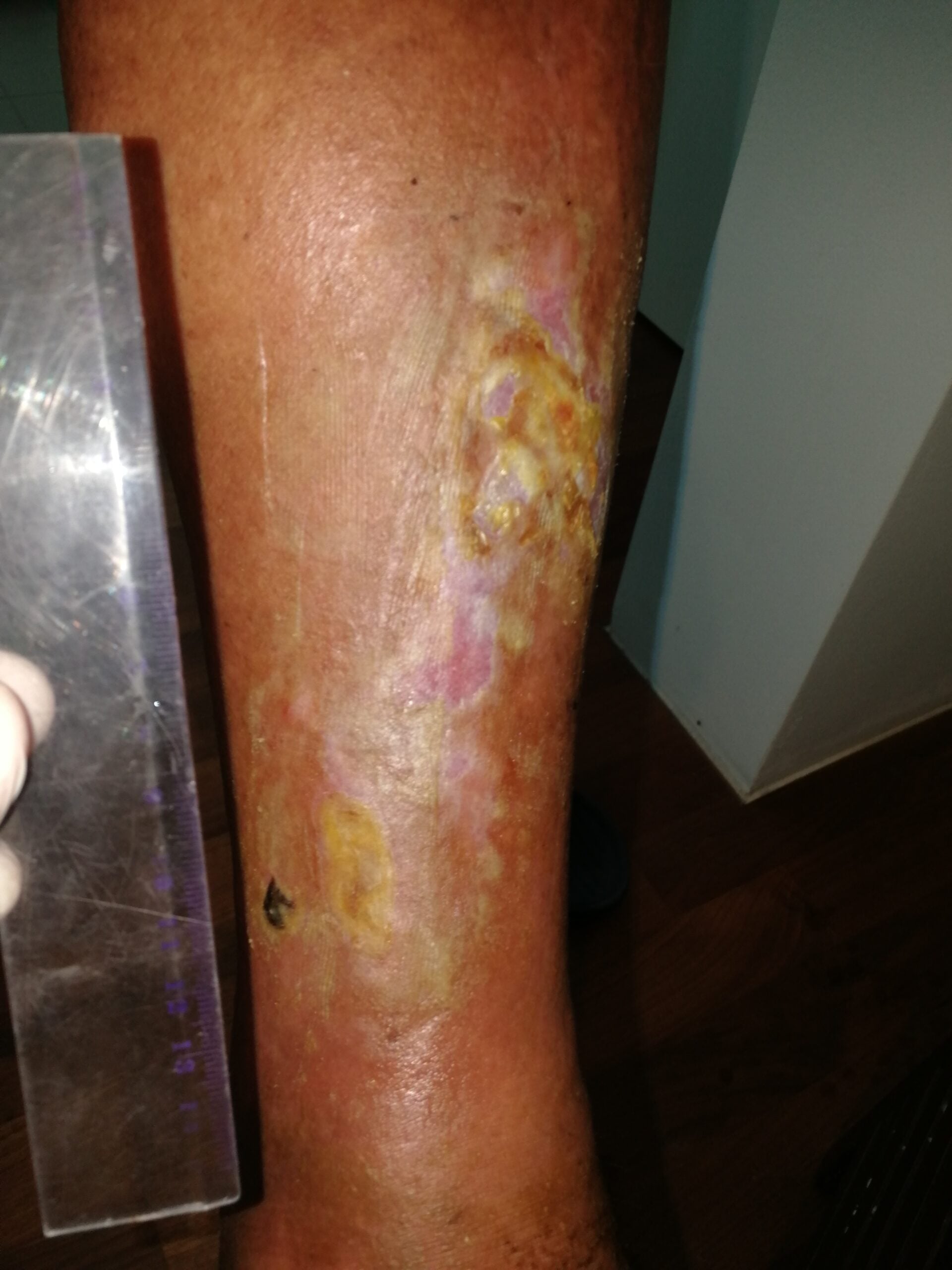
At the beginning of documented monitoring on 4 August 2018, the original wound measured 3.93 cm², while the secondary damaged area measured 21.15 cm² in total. The total estimated open wound area was therefore 25.08 cm².
How was the wound treated?
Documented monitoring during the use of Smrekovit Klasik began on 4 August 2018. The ointment was applied in an approximately 1–2 mm thick layer to sterile gauze, which was placed with the ointment-covered side directly onto the open wound and secured with a bandage.
The patient carried out the procedure at home twice daily. Approximately once a week, the wound was additionally examined, photographed and measured. This created consecutive time points at which changes in the open wound area could be compared.
When an individual wound was assessed as closed or epithelialised, Smrekovit Klasik was no longer used on that area. According to the original 2018 protocol, the patient then applied Smrekovit 365 to the closed skin without gauze or covering.
The procedure described above represents the actual documented wound care from 2018 and is not a current instruction for use. Current practical instructions for the use of Smrekovit Klasik on wounds are published separately.
How we measured the wound area
The area of the open part of the wound was estimated from serial photographs taken together with a measurement scale. The purpose of the measurements was to monitor changes in the open wound area over time as objectively as possible.
The photographs were graphically adjusted so that the scale was comparable between individual images. The boundaries of the open wound area were then marked manually and measured using a computer-based area measurement tool.
The original 2018 report already estimated a measurement uncertainty of approximately ±15%. The photographs were not taken at a fully standardised distance, angle or leg position, and the wound boundaries were determined manually.
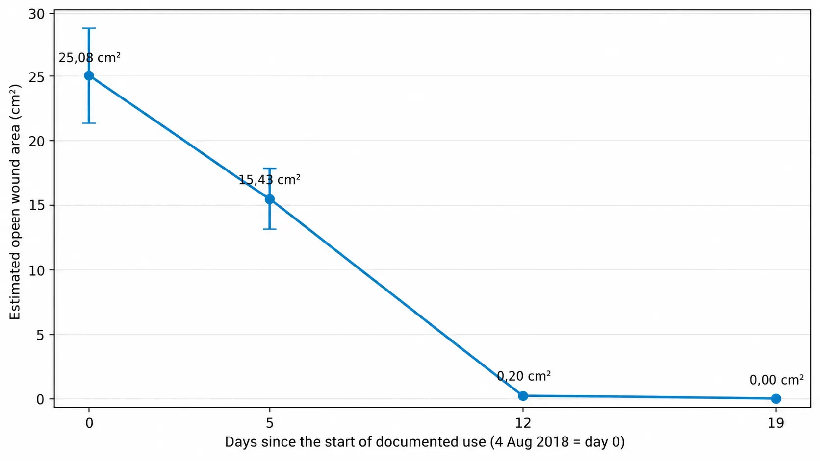
Despite this limitation, the serial measurements show a very clear trend: the total estimated open wound area decreased from 25.08 cm² at the beginning of monitoring to 15.43 cm² after five days, 0.20 cm² after twelve days and 0 cm² after nineteen days.
Photographic course of the wound
Open wound area reduced by approximately 38.5%
Five days after the start of documented monitoring, the total estimated open area of both wounds was 15.43 cm², compared with 25.08 cm² at baseline on 4 August.
The original post-traumatic wound measured approximately 0.81 cm², while the larger secondary damaged area measured approximately 14.62 cm². The total open wound area had therefore decreased by approximately 38.5% over five days.
At this examination, the patient also reported that the pain was only occasional and substantially less pronounced than at the beginning of monitoring.
From 25.08 cm², only 0.20 cm² of open wound area remained
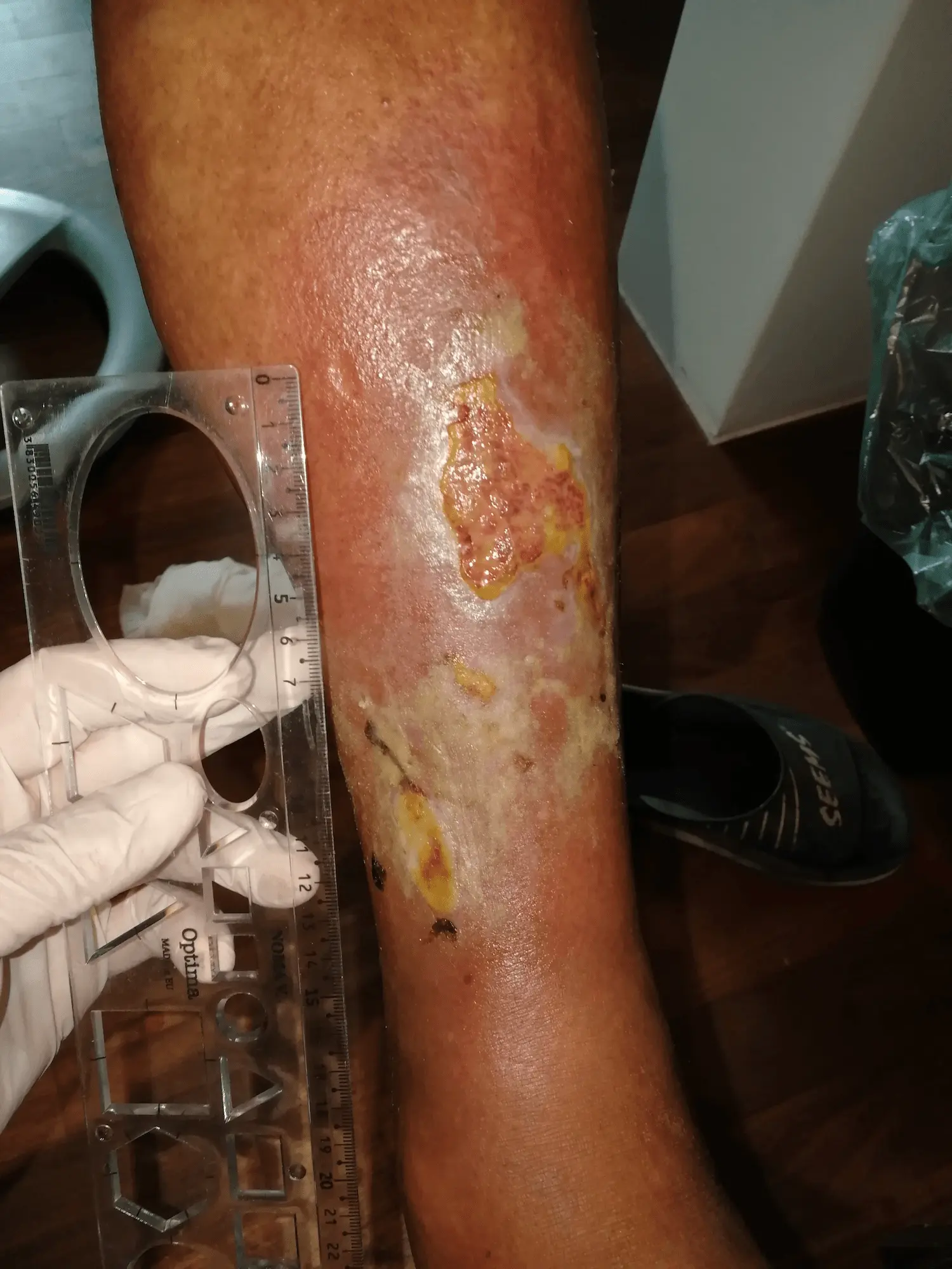
Twelve days after the start of documented monitoring, the original post-traumatic wound was assessed as closed. Only a small open area of the larger secondary damaged region remained, estimated at 0.20 cm².
Compared with the initial total open wound area of 25.08 cm², this represented an approximately 99.2% reduction in the estimated open wound area.
According to the record, the patient no longer reported pain at this examination. On the already closed area of the original wound, Smrekovit 365 was introduced according to the protocol used at the time, while the remaining open part of the wound continued to be treated with Smrekovit Klasik.
Both areas were considered closed at that time
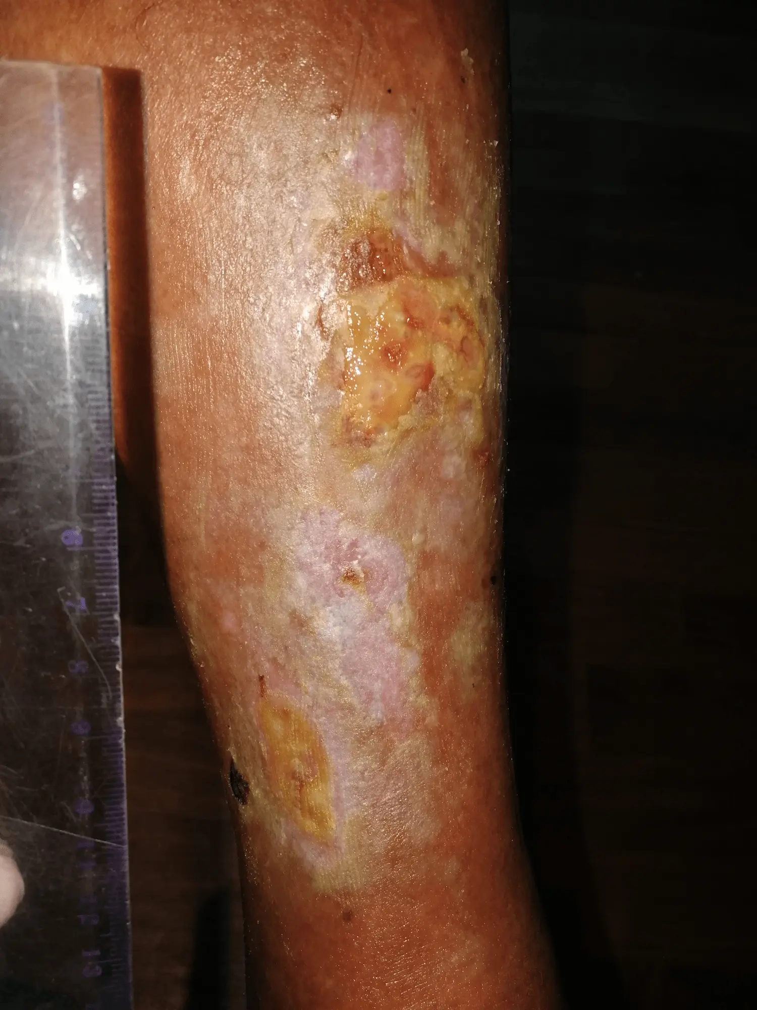
Nineteen days after the start of documented monitoring, the total estimated open wound area was 0 cm². Both wounds were considered epithelialised at that time.
The estimated total open wound area had therefore decreased from the initial 25.08 cm² to 0 cm². Smrekovit Klasik was no longer used on the open wound from that day onward.
According to the 2018 protocol, the patient continued local application of Smrekovit 365 to the closed skin without gauze or covering.
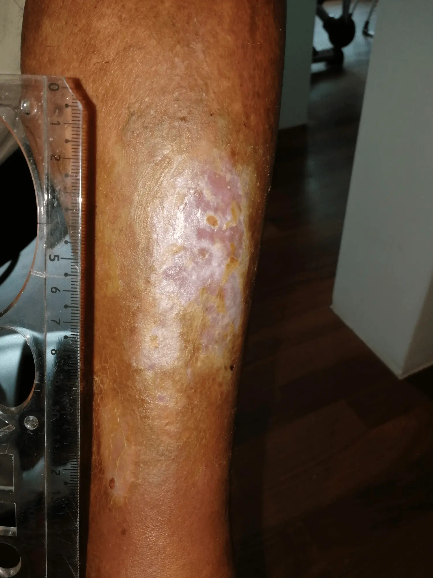
Follow-up photograph after wound closure
Nine days after both areas had been assessed as epithelialised, the open wound was no longer clearly visible in the follow-up photograph.
At the sites of the former wounds, pink, yellowish-brown and pigmented skin changes were still present, consistent with continued remodelling of the new tissue after wound closure.
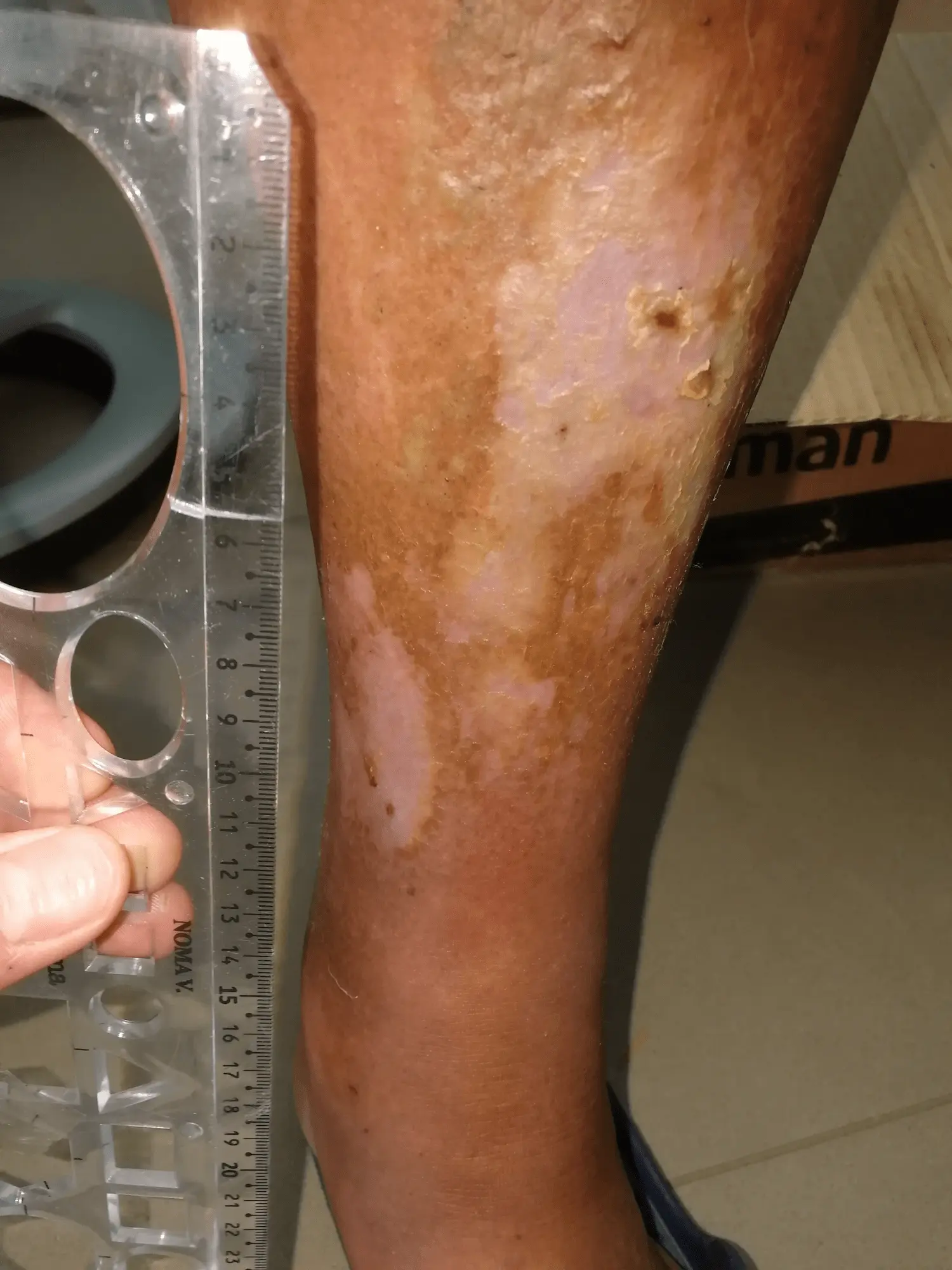
The area remains closed
In the last preserved follow-up photograph, the open wound is no longer clearly visible. The area remained closed, with no reopening of the wound.
Residual colour and texture changes of the skin are still present, together with a small superficial irregularity in the upper part, but the photographic course confirms that the area remained closed.
What is important about this case?
The greatest value of this case lies in the fact that the course was documented not only descriptively, but also with serial photographs and measurements of the open wound area. This allows substantially more precise monitoring of changes over time than a typical user experience.
At the beginning of documented monitoring, the total estimated open wound area was 25.08 cm². After five days it was estimated at 15.43 cm², after twelve days at 0.20 cm², and after nineteen days the open wound area was no longer measurable.
The patient’s age and the presence of venous and heart failure also provide important context. The severity of both conditions is not objectively defined in the preserved documentation, so their effect on the rate of healing cannot be assessed precisely.
It is also important that documented monitoring did not begin immediately after the injury. The patient had already been receiving medical care for approximately two weeks, and documented use of Smrekovit Klasik began 16 days after the injury.
The course is very clearly associated in time with the period of regular use of Smrekovit Klasik, but a single retrospective case without a control group does not allow the conclusion that the product was the sole or necessary cause of wound closure.
Documentation and limitations of the case
The case was reconstructed retrospectively from several preserved sources: the original 2018 report, a brief questionnaire, dated photographs, measurement appendices and wound-area calculations.
The strongest part of the documentation consists of the serial photographs with a measurement scale and the calculated values of the open wound area. The measurements were made from non-standardised photographs with manually defined wound boundaries, so the original report already estimated a measurement uncertainty of approximately ±15%.
The original medical reports and records of the preceding care are not among the preserved sources. Information about the medical visit, regular dressing changes, and venous and heart failure is therefore based on the preserved questionnaire, the original report and the case data. The severity of the accompanying conditions cannot be defined more precisely.
It also cannot be reliably confirmed that the polyurethane dressing used previously caused the larger secondary damaged area. The temporal sequence is documented, but a causal relationship has not been established.
This is a single documented observational case without a control group. It therefore cannot establish the causal effectiveness of any individual intervention or predict the same course in other wounds.