
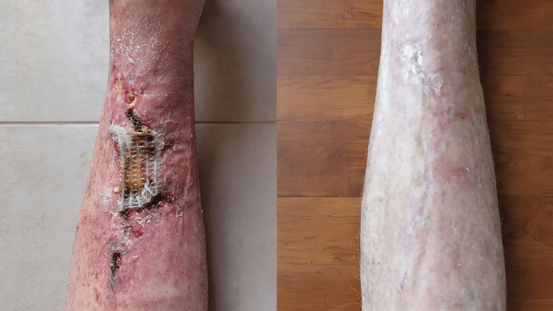
Case in brief
- Patient
- Male, 92 years old at the start of follow-up
- Start of documented follow-up
- Problem
- Chronic lower-leg ulcer, with a course consistent with a venous leg ulcer
- Duration before Smrekovit Klasik
- According to the family, about one and a half months
- Previous care
- Saline solution, ointment according to medical instructions and later zinc dressings
- Important associated factors
- Heart failure, treatment with Marevan and borderline elevated blood sugar
- Use of Smrekovit Klasik
- On sterile non-woven gauze, with regular dressing changes and photographic monitoring
- Final status
- – the wound was practically closed, with epithelialisation of the area
- Documentation
- Time-stamped written correspondence, serial photographic monitoring and an anonymised public case report
Smrekovit Klasik was used as part of the wound care. This case shows the documented course of one chronic lower-leg ulcer and does not represent a clinical study or proof of the isolated effect of one intervention.
Table of contents
Baseline condition and previous care
The first photograph of the wound was sent on 2 March 2026. The case involved a 92-year-old male patient with a chronic wound on the lower leg. According to the family, the wound had been open for about one and a half months before Smrekovit Klasik was started.
Before that, the wound had been treated according to medical instructions. The visiting nurse cleaned it with saline solution and applied an ointment, the exact name of which had not been finally confirmed at the time this report was prepared. According to the family, this care did not lead to improvement.
A dermatology review was also carried out during this period. Zinc dressings were applied, after which the condition reportedly worsened. The patient had confirmed heart failure, was treated with Marevan, and his blood sugar was described as borderline elevated.
Based on its appearance, location and course, the wound was consistent with a chronic lower-leg ulcer of venous origin. However, because the full medical record was not used in this public report, this is stated as a clinical or observational interpretation based on the course, photographs and family-provided information.
Use of Smrekovit Klasik and wound monitoring
From the start of documented follow-up, Smrekovit Klasik was used on sterile non-woven gauze. The gauze was placed directly onto the wound with the ointment-covered side facing the wound, and dressing changes were adjusted according to the wound condition and the amount of exudate.
Follow-up was carried out remotely, mainly through time-stamped messages and serial photographs. The family regularly reported on pain, exudate, the appearance of wound coverings, odour, bleeding and the gradual closing of the wound surface.
In this type of follow-up, it is important not to interpret every change too quickly. Yellowish coverings were treated as part of the wound process in this case, while mechanical removal was not recommended. If signs of infection, rapid deterioration, strong odour, fever or a marked increase in pain appear, medical review is always appropriate.
The detailed practical procedure for open wounds is described separately in the instructions for wound care. This case shows how that approach was used in one concrete, documented course of a chronic lower-leg ulcer.
Photographic course of the wound
Chronic lower-leg ulcer at the start of follow-up
The first available photograph shows a chronic wound on the lower leg of a 92-year-old male patient. According to the family, the wound had already been open for about one and a half months at that time.
Before Smrekovit Klasik was started, the wound had been treated according to medical instructions, including saline solution and an ointment, and later also zinc dressings after a dermatology review.
The photograph shows a chronic ulcer with surface coverings and irritated surrounding skin. Based on its location, appearance and associated factors, the course was consistent with a lower-leg ulcer in the context of impaired circulation or venous insufficiency.
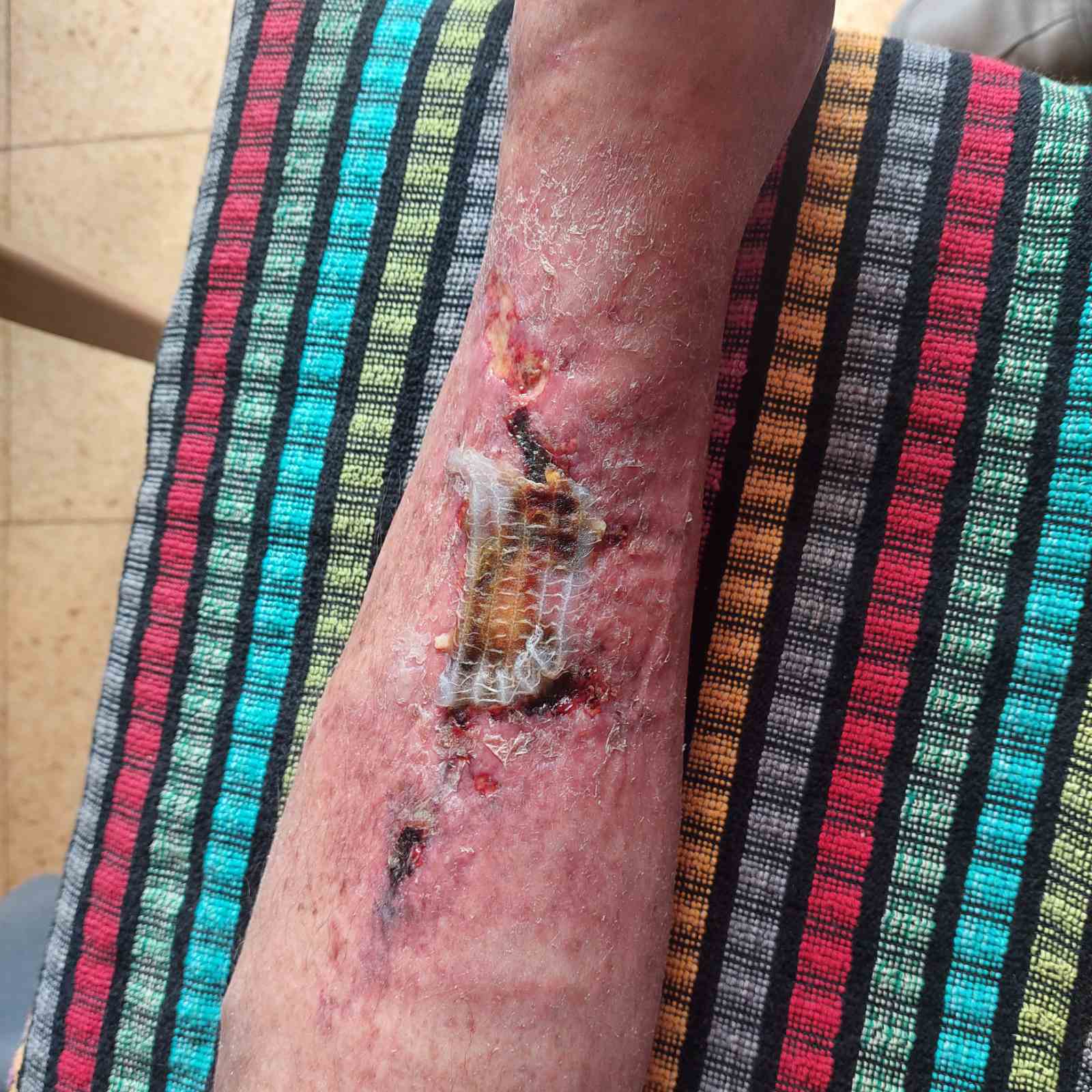
2 March 2026 – initial state at the start of documented follow-up; according to the family, the wound had already been open for about one and a half months.
Yellowish coverings and mild odour caused concern
Twenty-five days after the start of documented follow-up, the family expressed concern about yellowish coverings on the wound and a mild odour. In chronic wounds, this type of appearance often creates uncertainty because it is difficult to distinguish from signs of deterioration based on a photograph alone.
In the communication, it was emphasised that the yellowish coverings should not be removed mechanically unless there was a clear professional reason to do so. At the same time, it was important to monitor whether the odour increased, or whether marked redness, warmth, increased exudate, fever or rapid deterioration of the general condition appeared.
This point is important because it shows one of the typical dilemmas in remote follow-up of a chronic wound: some changes may be part of the wound process, while signs of infection or deterioration still need careful attention.
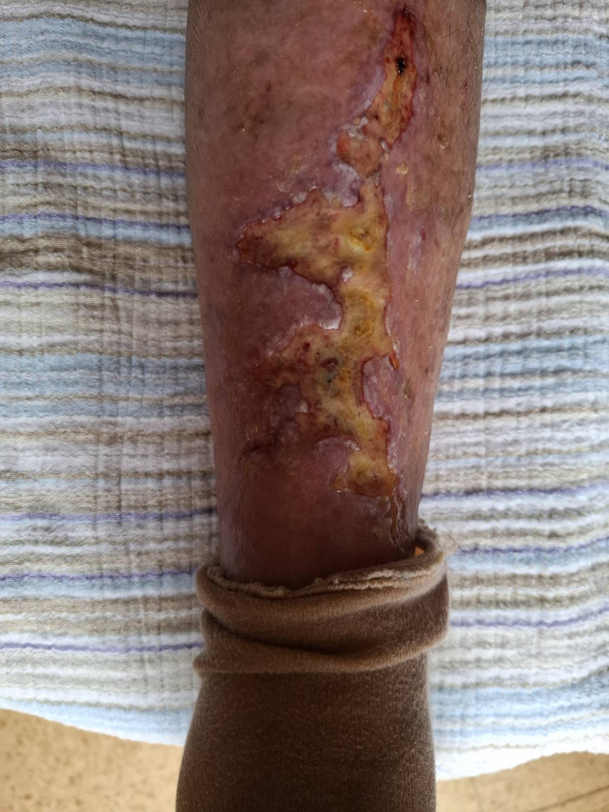
27 March 2026 – the photograph shows a phase with yellowish coverings and mild odour, which led the family to express concern about the wound course.
Visible signs of epithelialisation and the question of continuing care
In early April, the family considered whether it might be better to let the wound “dry out”. In the communication, it was advised not to stop the care too early, as the photograph already showed signs of gradual surface closure and formation of new skin.
The wound was still in the healing process, but the affected area appeared calmer than during the first weeks. It was important not to dry out the open part too early and to protect the newly forming, sensitive surface.
This point is important because it marks the transition from a clearly active chronic wound phase to a stage where gradual epithelialisation and surface closure could already be seen.
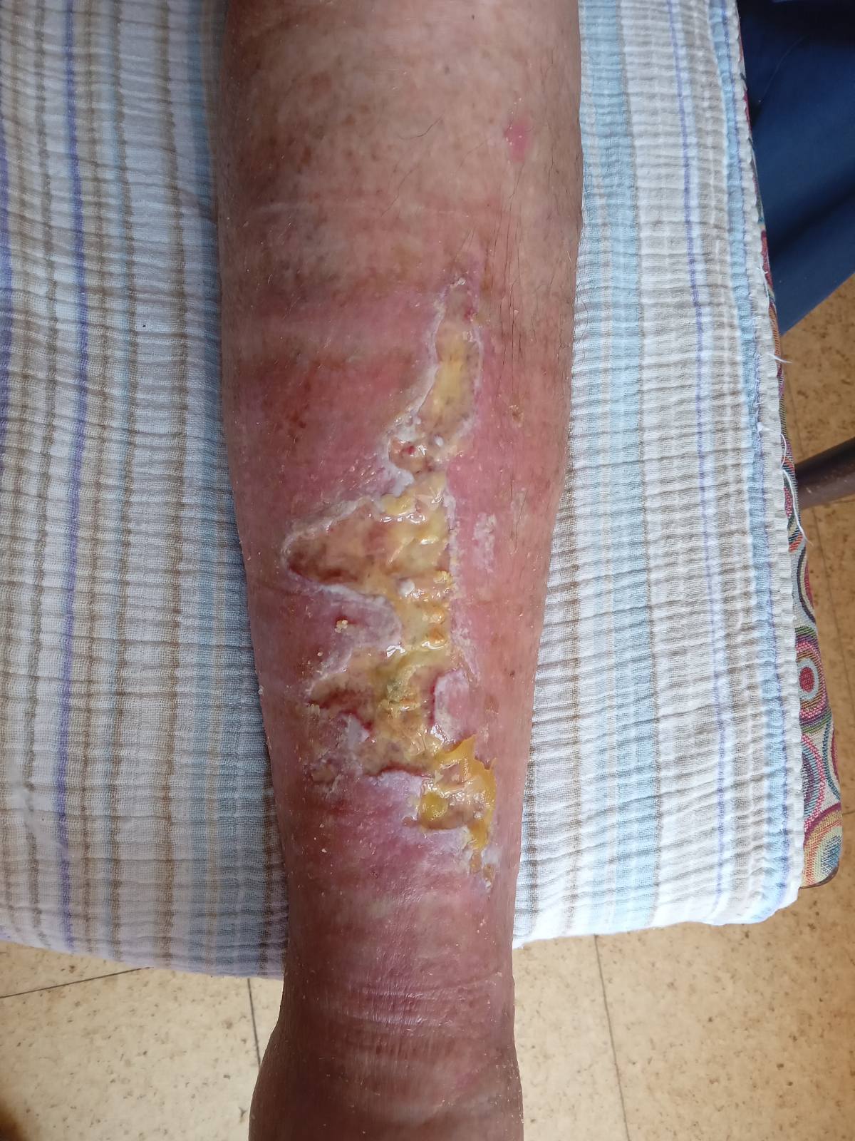
9 April 2026 – the affected area showed signs of gradual surface closure and formation of new skin.
Clearer change: fewer coverings, more granulation and less pain
By late May, a clearer shift was visible compared with previous follow-ups. There were fewer yellowish coverings, and a larger part of the wound showed the appearance of more active granulation tissue.
During this period, the family reported that pain was still present, but it was more occasional and milder. This was important because in previous weeks the patient had repeatedly described pain as a significant part of the problem.
This checkpoint is important because, after a slow early course, a more visible change in the wound appearance became apparent: fewer coverings, more granulation and a clearer transition towards closure of the affected area.
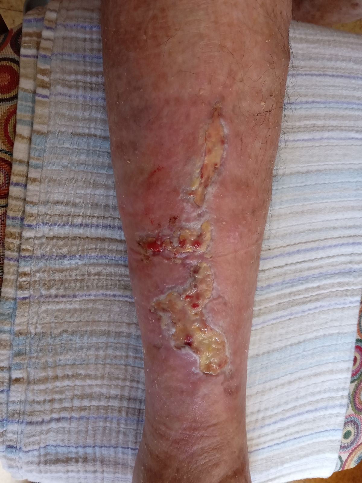
27 May 2026 – compared with previous follow-ups, fewer coverings, more granulation tissue and a clearer transition towards wound closure were visible.
Most of the wound had already closed, while the lower part remained the most active
By the second half of June, the healing course had become much more evident. Larger parts of the affected area were already covered with new skin, while the lower part of the wound remained open or more active.
During this period, the family reported that the condition was visibly better, and the patient was satisfied with the progress. Nevertheless, the wound was not yet completely closed, so it remained appropriate to continue protecting the sensitive area and monitoring the lower part of the wound.
This point is important because it shows the transition from an active granulation phase to a stage where most of the surface had already epithelialised, while the remaining open part still required further care.
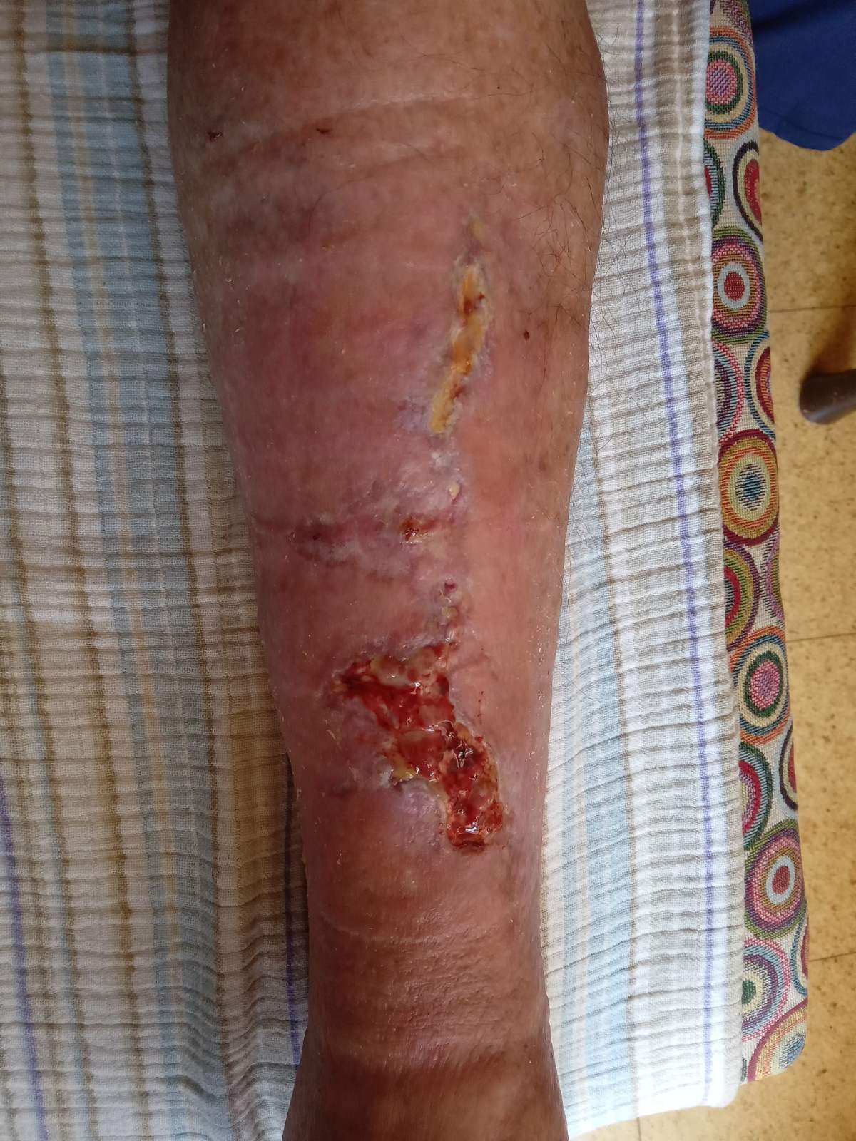
20 June 2026 – most of the affected area was already covered with new skin, while the lower part of the wound remained the most active.
The main wound was almost closed
In early July, the main lower-leg wound was almost completely closed. Most of the affected area was covered with new skin, while only a smaller part remained open or more sensitive.
At this stage, it was important to continue protecting the sensitive area, as newly formed skin is not yet as resilient as mature skin. In chronic wounds, even after near-complete closure, it may still be appropriate to monitor whether the surface reopens, becomes irritated or produces exudate.
This point is important because it shows the transition from active healing to the final phase of epithelialisation, when the wound no longer appeared as an extensive open ulcer, but the process was not yet fully complete.
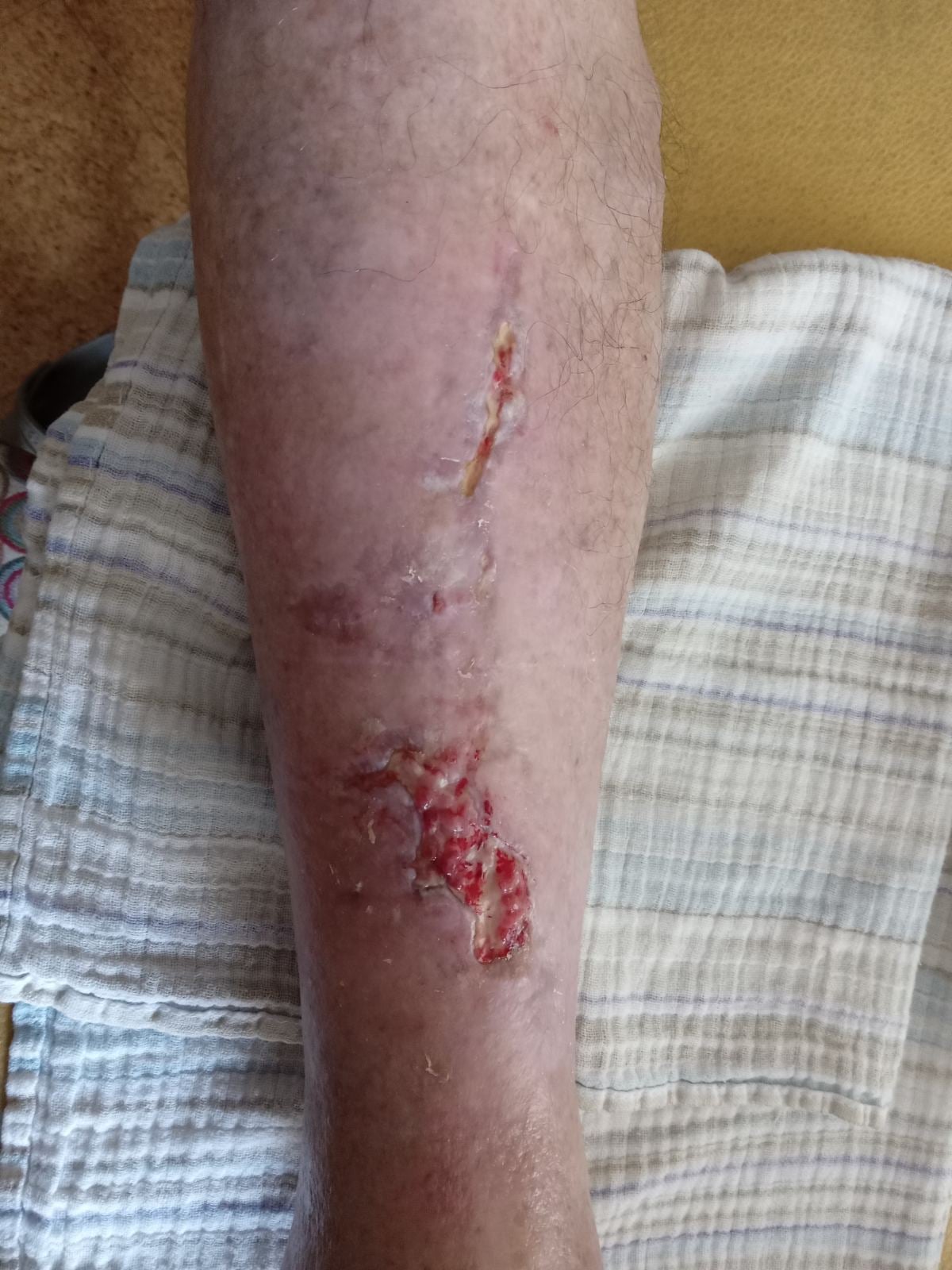
3 July 2026 – the main wound was almost completely closed, with only a smaller part still open or sensitive.
The wound was almost completely closed
By early August, the main lower-leg wound was almost completely closed. The affected area no longer had the appearance of an extensive open ulcer, but mainly showed sensitive new skin and residual changes after a prolonged healing process.
At this stage, the main focus was to continue gentle protection of the skin and monitor whether any smaller sensitive areas reopened. After such a long chronic wound course, newly formed skin remains fragile and less resistant for some time.
This point is important because it shows that by early August the case had moved from the phase of an open chronic ulcer into a stage of near-complete closure, although it was still not appropriate to declare a fully final state.
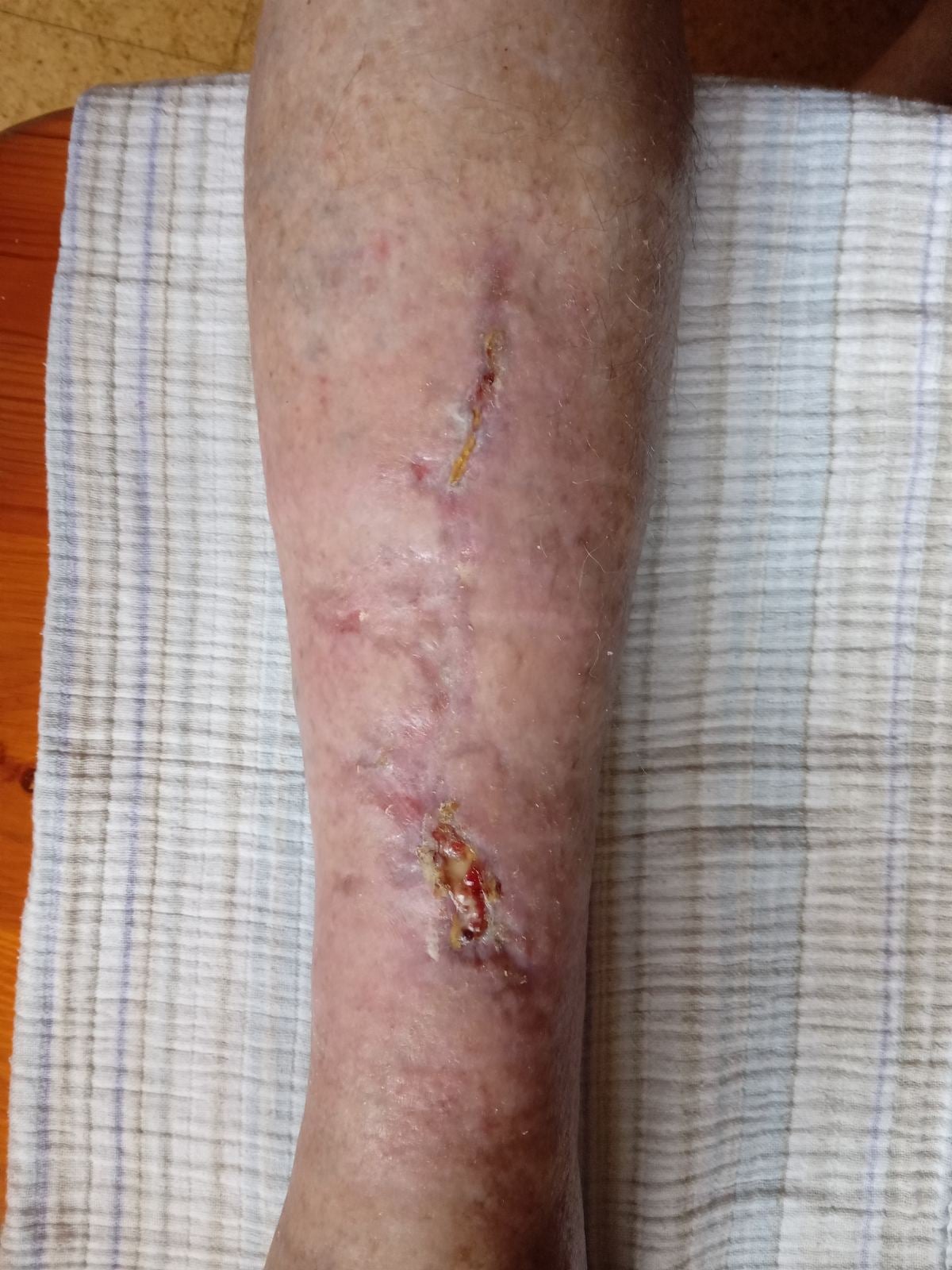
3 August 2026 – the main wound was almost completely closed; the image mainly showed sensitive new skin after a prolonged healing process.
Final follow-up: the wound was practically closed
The final photograph at the end of September shows that the open wound was no longer clearly visible. At the site of the previous ulcer, mainly skin changes, scarring and sensitive new skin remained after a prolonged healing process.
Over several months of follow-up, a transition was documented from a chronic open lower-leg ulcer to a state that was practically closed or epithelialised. At the same time, the case shows that the course was slow, with periods of pain, coverings, exudate, uncertainty and gradual surface closure.
This final point does not prove the isolated effect of one intervention, but it clearly documents the course of one chronic lower-leg ulcer during the period of Smrekovit Klasik use and regular wound care.
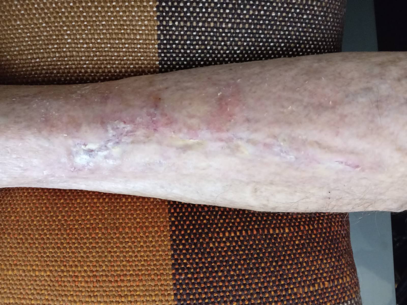
29 September 2026 – final follow-up photograph; the open wound was no longer clearly visible, with mainly residual skin changes after prolonged healing.
The other leg and early skin changes
During follow-up of the main wound, early skin changes also appeared on the other leg. The family photographed them on 20 June and in early July 2026, because their appearance reminded them of the beginning of a similar process to the one that had previously developed on the main wound.
Smrekovit Klasik was used on these areas early, before a larger open wound could develop. According to later messages, these areas did not open into a larger ulcer and did not progress in the same way as the original wound on the other lower leg.
This part of the case cannot be interpreted as proof that a new ulcer was prevented, because there was no control comparison and the early changes could have settled for other reasons as well. It is nevertheless a practical observation, as it shows that after the experience with the main wound, the family began to monitor similar changes earlier and more carefully.
What the case shows and what it does not show
This case clearly shows that in a 92-year-old patient with a chronic lower-leg ulcer, a several-month course was documented from an open, painful wound with surface coverings to a state where the open wound was no longer clearly visible. The course was followed with photographs and time-stamped written communication with the family.
The case also shows that healing was not linear. There were periods of stronger pain, concern about yellowish coverings, mild odour, slow reduction of the open surface, granulation and gradual formation of new skin. In chronic wounds, this is important because a single follow-up photograph does not tell the whole story by itself.
However, the case does not prove that Smrekovit Klasik independently caused the wound closure. It was an individual observational case, without a control group and without the possibility of reliably separating the effect of the product from other factors, such as regular care, the patient’s general condition, reduced strain on the leg and the natural course of the wound.
The most accurate way to understand this case is therefore as a documented course of a chronic lower-leg ulcer during the period of Smrekovit Klasik use, not as clinical proof of a therapeutic effect.
Documentation and limitations of the case
The case is based on time-stamped written communication with the family, a series of wound photographs, subsequently obtained information about previous care, and an anonymised public case report. The photographs provide a useful view of the visual wound course, but they were not taken under standardised conditions.
An important limitation is that the public description is not based on a full review of the medical records. Information about previous care, wound duration, pain, the patient’s response and observations by visiting nursing care therefore partly relies on statements from the family.
There was also no control group, no standardised wound surface measurements and no independent clinical assessment of the effect of any single intervention. The case therefore cannot show what the course would have been without Smrekovit Klasik or what the result would be in other similar wounds.
Because the author or preparer of the report is connected with Smrekovit, this represents a potential conflict of interest and a possible source of interpretative bias. For that reason, cautious wording is used to distinguish the documented course from conclusions about causality.