
Case in brief
- Patient
- 44 years old at the time of injury
- Date of injury
- Injury
- Dislocation of the right ankle joint after landing
- Emergency treatment
- Joint reduction followed by X-ray imaging; no associated fracture is described in the available documentation
- Initial immobilization
- Non-weight-bearing plaster splint, later replaced at the patient’s own decision with a removable Vacoped system
- Use of Smrekovit Klasik
- Smrekovit Klasik with a compress under the removable support system; the compress was worn practically continuously and additional ointment was applied approximately once daily
- First steps without the brace
- Approximately one week after the injury
- Walking without a limp
- Approximately two weeks after the injury, the patient reported walking around 1500 m without a limp
- After approximately one month
- Several kilometres of walking per day and short running intervals of a few dozen metres
- Documentation
- Emergency medical report, X-rays after reduction and follow-up images, serial dated photographs and a subsequent written reconstruction of the functional course
The case is of interest because of the early functional recovery after a documented ankle dislocation that required urgent reduction. The course of recovery coincided in time with the use of Smrekovit Klasik with a compress, but this is a single retrospectively documented observational case, so no causal effectiveness of any individual intervention can be determined from it.
Table of contents
How did the dislocation occur?
The injury occurred on 9 November 2018, when a 44-year-old man injured his right ankle after a rapid jump or landing. Because of the injury, he was transported by ambulance to the emergency department.
On examination, the right ankle joint was visibly deformed, and the skin over the joint was tense. The pulse on the dorsum of the foot was palpable, while the patient was otherwise described as stable on arrival.
The clinical picture was consistent with a dislocation of the right ankle joint. The joint therefore first had to be urgently returned to its anatomical position – a reduction was performed.
An important feature of the case is that the available documentation does not describe an associated fracture. Fractures are often associated with this type of injury, so according to the patient’s later account, he was initially also prepared for the possibility of surgical treatment.
What was done in the emergency department?
How did home care proceed?
After discharge from the emergency department, the patient decided on his own to remove the plaster splint and replace it with a removable immobilization boot, Vacoped.
Under the boot, he used a compress with Smrekovit Klasik. According to the original report, the compress was worn practically continuously, during the day and at night, and the ointment was reapplied to the compress approximately once daily.
According to his later account, the patient walked with the immobilization boot from the beginning. He tried his first cautious and controlled steps without the boot approximately one week after the injury, after which he used it again for protection against a possible misstep or fall.
Approximately two weeks after the injury, according to his own report, he stopped using the boot. At that point, he was already walking without it, but still cautiously and with additional ankle bandaging.
Removal of the prescribed plaster splint and the transition to removable immobilization were the patient’s own decisions. This course is described here as part of the documentation of this specific case and does not represent a recommendation for the treatment of ankle dislocation.
Photographic course of the ankle
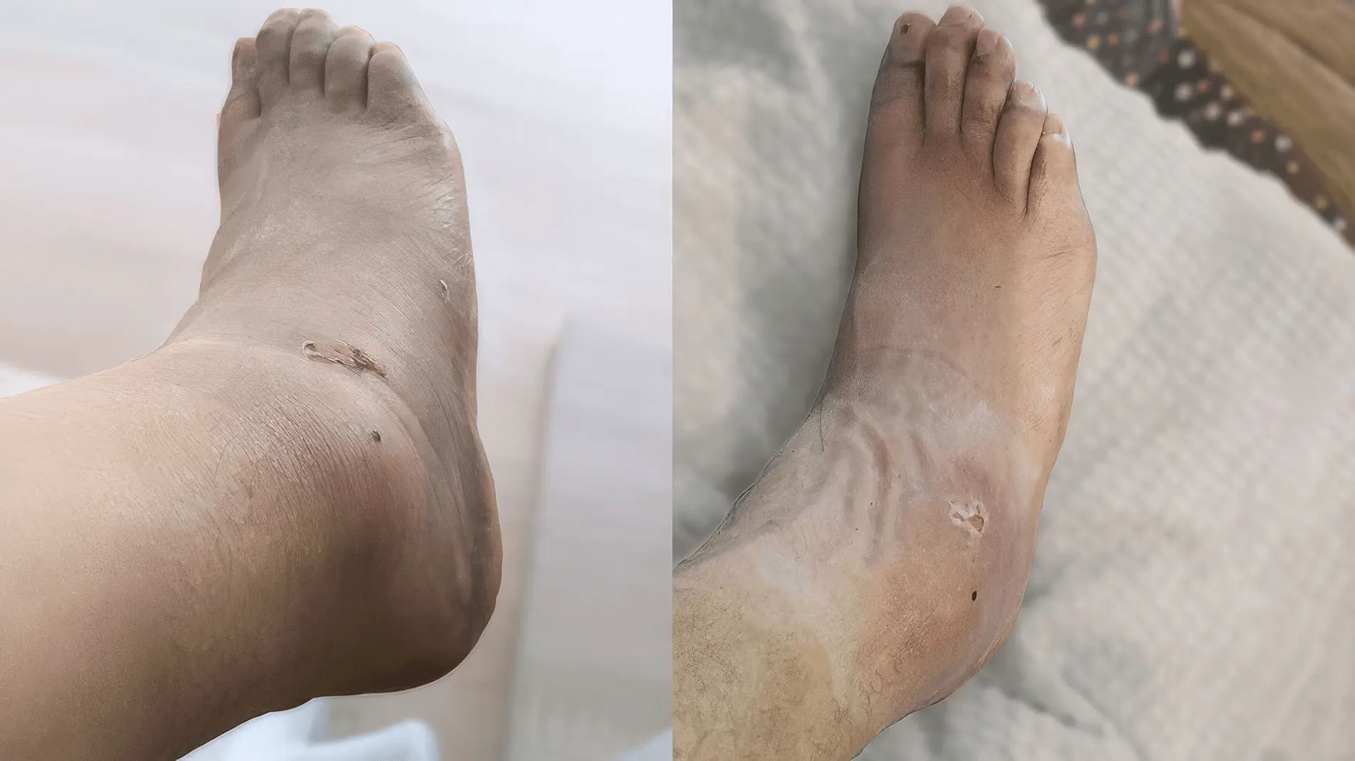
From the fifth day after the injury, a series of dated photographs has been preserved, allowing the changes in swelling, bruising and the appearance of the soft tissues of the right ankle to be followed.
The photographs were not taken according to a standardized protocol, so they do not allow precise measurement of the extent of swelling. However, the sequence does allow a visual comparison of the course over approximately one month after the injury.
Marked swelling and bruising
Five days after the dislocation, the photographs still show marked swelling of the ankle and foot as well as extensive bruising. A superficial skin injury is also present in the ankle area.
This is the first preserved photographic time point, so we use it as the baseline for subsequent visual comparison.
The swelling is visibly less pronounced
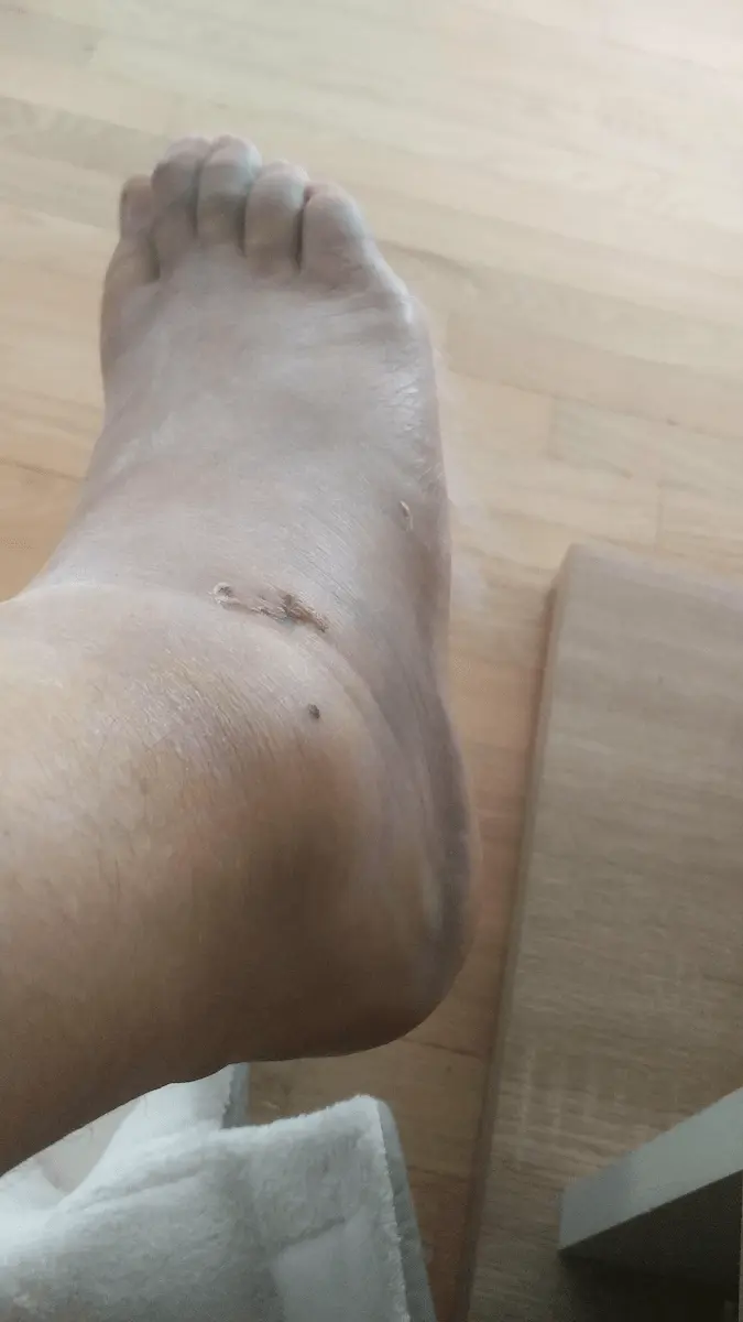
On the twelfth day after the injury, the ankle is still swollen in the photographs, but the swelling is clearly less pronounced than on day five. The extent of visible bruising is also decreasing.
During this period, the patient was already taking cautious steps without the immobilization boot, although he still used it for protection.
Only minor residual swelling remains
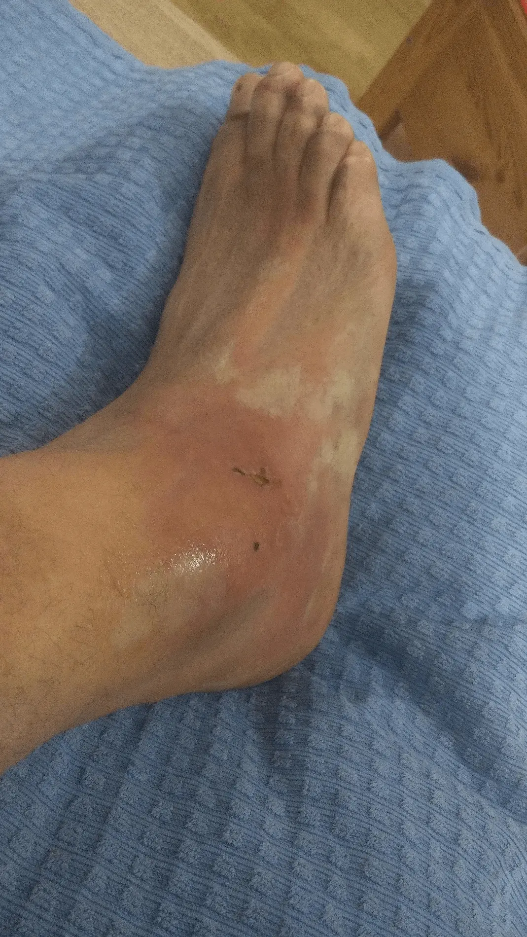
Fifteen days after the injury, the shape of the ankle in the photograph is already much closer to its usual appearance. Some residual swelling and skin discoloration are still present, but both are much less pronounced than in the first images.
At around this time, according to the later written reconstruction, the patient had already stopped regularly using the immobilization boot and completed a walk of approximately 1500 metres without limping. This functional information is based on the patient’s report and was not objectively measured.
The appearance of the ankle is approaching normal
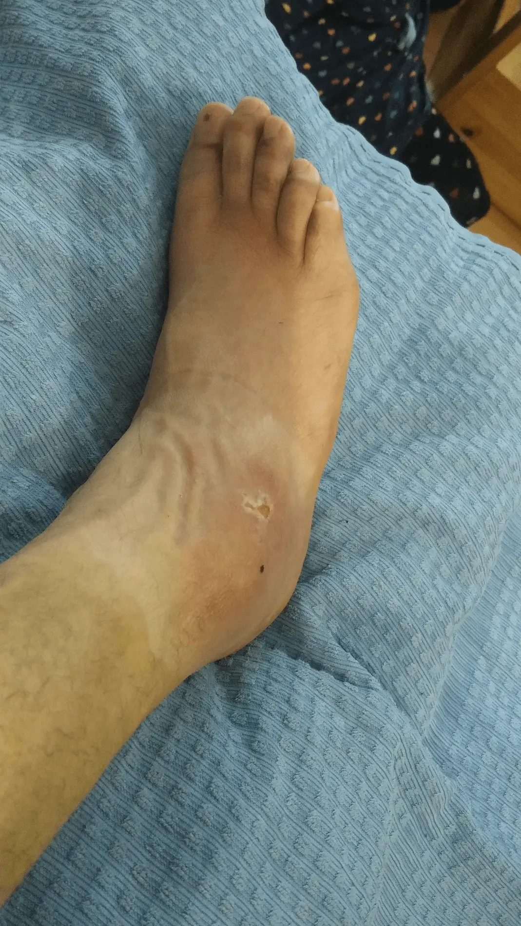
Just under a month after the injury, pronounced swelling is no longer visible in the photograph. The contours of the ankle are clearly visible, while residual skin discoloration remains in areas affected by the previous bruising and superficial injury.
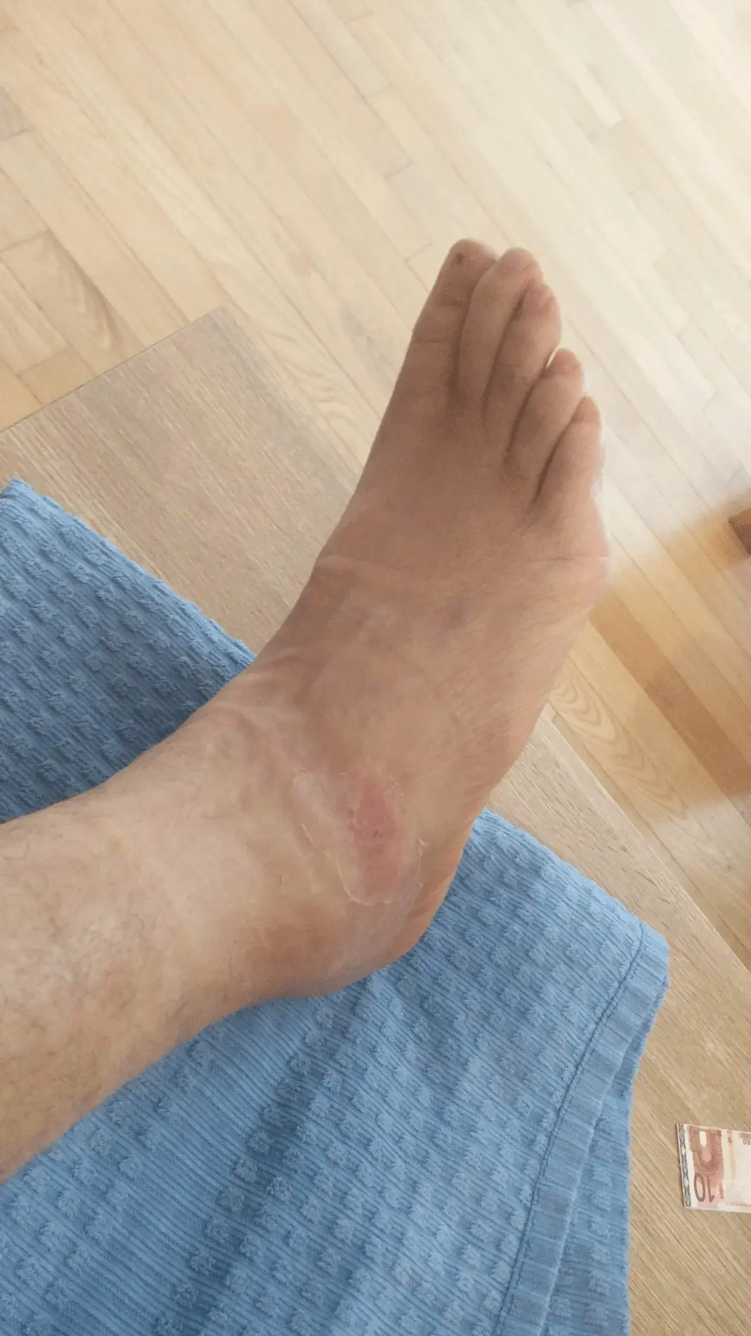
Last preserved photographic follow-up
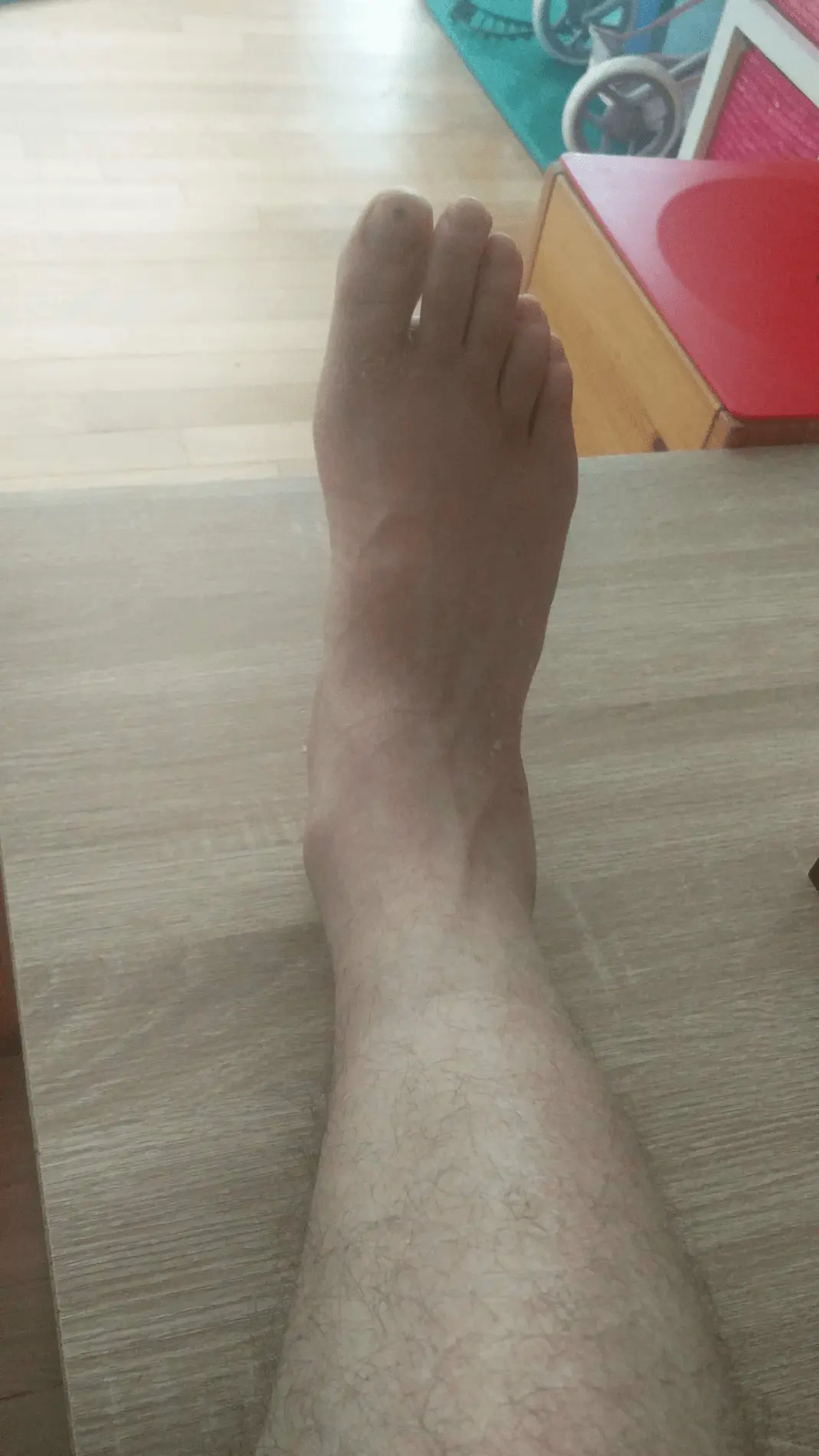
Five weeks after the injury, the last preserved photographs show that pronounced swelling is no longer visible. The shape of the ankle appears normalized, while minor residual changes in skin colour are still present.
The photographs themselves do not allow an assessment of stability, mobility or ligament healing. The functional course is therefore documented separately on the basis of the patient’s written statements.
How quickly did ankle function return?
In addition to the photographic follow-up, the patient’s later written reconstruction of the return to walking and loading of the injured ankle has also been preserved. The functional milestones therefore rely mainly on the patient’s own report and were not verified using standardized functional tests.
- Approximately 1 week
- First cautious and controlled steps without the immobilization boot. After trying this, the patient resumed using it mainly for protection against a misstep or fall.
- Approximately 2 weeks
- The patient stopped regularly using the immobilization boot and, with additional bandaging, completed a walk of approximately 1500 metres without limping.
- Approximately 1 month
- According to his own report, he was already walking several kilometres per day without pain.
- Approximately 1 month
- He also began incorporating short running intervals of several dozen metres into his walking.
Considering the initial condition, when the dislocation required urgent reduction and non-weight-bearing immobilization was prescribed at discharge, the described milestones indicate an early return to functional loading of the ankle.
The functional milestones describe the ability to walk and bear weight, but not necessarily complete anatomical healing of the injured structures. The preserved documentation contains no standardized measurements of mobility, stability, strength or pain, and no soft-tissue imaging was performed that could have more precisely defined the extent of the ligament injury and its healing.
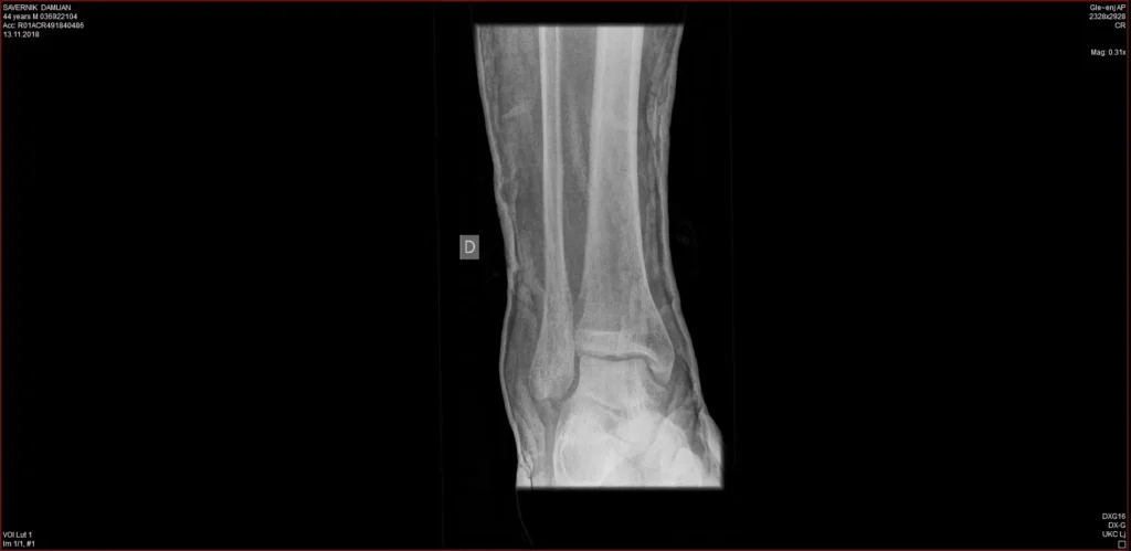
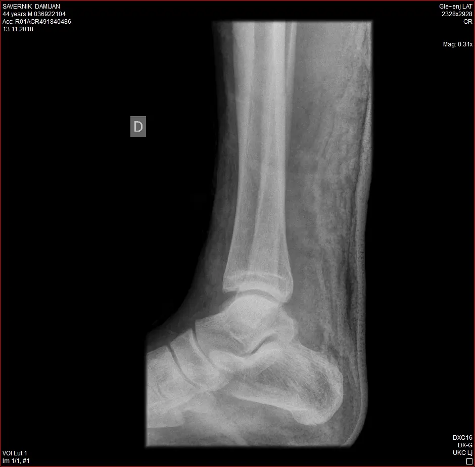
X-rays after reduction
X-ray imaging was performed on 9 November 2018 after reduction of the right ankle joint. The emergency report states that after reduction the position of the ankle was anatomical.
No associated fracture is described in the available documentation. Follow-up X-rays from 13 November 2018, four days after the injury, have also been preserved.
The images are shown here as part of the case documentation and not as an independent radiological interpretation. They also do not allow assessment of the extent of ligament injury or ligament healing.
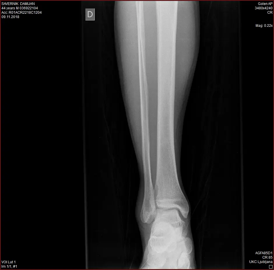
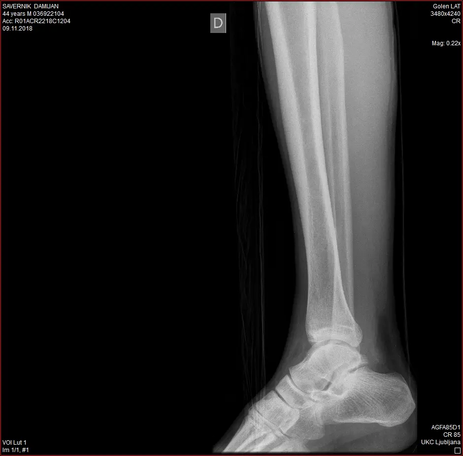
The emergency report accompanying these images states that the ankle was in anatomical alignment after reduction.
What is important about this case?
The case is particularly interesting because this was not a mild sprain, but a dislocation of the right ankle joint that required urgent reduction. After reduction, the ankle was initially immobilized with a non-weight-bearing plaster splint.
Read more about what happens to the ligaments in an ankle sprain, how injuries differ in severity and how recovery usually progresses in the article Ankle sprain: symptoms and recovery. If you are primarily interested in the practical use of Smrekovit products for a sprain, the instructions are collected in the section Use for ankle sprain.
Despite the initial severity of the injury, according to the patient’s later written report, he took his first cautious steps without the immobilization boot approximately one week after the injury, and about two weeks after the injury he walked around 1500 metres without limping.
Approximately one month after the injury, according to his own report, he was already walking several kilometres per day without pain and was also incorporating short running intervals of several dozen metres into his walking. The photographic series from the same period also shows a gradual reduction in swelling and bruising and a return towards the ankle’s usual appearance.
For context, comparison with the published literature is also informative. A systematic review of 154 reported cases of ankle dislocation without an associated fracture found an average immobilization period of slightly more than six weeks, with a range of 2 to 16 weeks. In this context, discontinuing regular use of the immobilization boot approximately two weeks after the injury was early in this case. Direct comparison between individual cases is not possible, however, because of differences in the injuries and treatment.
This entire course coincided with the use of Smrekovit Klasik with a compress under the removable immobilization boot. However, a single retrospectively documented case does not allow us to determine how much of the observed course can be attributed to the product and how much to natural healing, reduction, immobilization and gradual loading of the joint.
The particular value of this case therefore lies mainly in the combination of medical documentation, X-rays, serial photographs and function-related milestones reconstructed in writing, which allows the course after the injury to be followed relatively well.
Documentation and limitations of the case
The case was retrospectively reconstructed from several preserved sources: the emergency report, X-rays taken after reduction and at the follow-up examination, serial dated photographs, the original case report and later written correspondence with the patient.
The strongest part of the documentation consists of the medical record of the reduction and immobilization, the preserved X-rays and the photographic series, which allows changes in the appearance of the ankle to be followed over time.
Functional milestones, such as the first steps without the immobilization boot, approximately 1500 metres of walking without limping and later walking several kilometres per day, are based on the patient’s later written reconstruction. These data were not verified using standardized functional tests.
No X-ray taken before reduction is preserved in the available documentation, so the original direction of the dislocation cannot be reliably reconstructed. No soft-tissue imaging, such as magnetic resonance imaging, was performed either, which could have defined the extent of the ligament injury more precisely.
No standardized measurements of swelling, range of motion, stability, strength or pain have been preserved. The photographs therefore allow mainly the visual course of swelling, bruising and soft-tissue changes to be followed, but not anatomical ligament healing or complete functional normalization of the joint.
Another important uncontrolled factor is the patient’s decision to remove the prescribed plaster splint after discharge and replace it with a removable immobilization boot. The course of recovery may also have been influenced by reduction, immobilization, gradual loading, the natural healing process and other factors that cannot be separated from one another in an individual case.
This is a single retrospectively documented observational case without a control group. It therefore cannot be used to determine the causal effectiveness of any individual intervention or to predict the same course in other injuries.